Biology Reference
In-Depth Information
a
b
ap
h
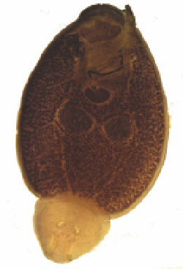
Figure 8.2
(a) Light micrograph of the marine parasite
(ventral view). Anterior pads (ap) bind to fish skin using a
temporary adhesive secretion, and the haptor (h) attaches
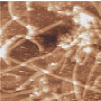
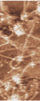
to host skin by suction (scale bar, 1 mm). (b) Atomic force
microscopy (AFM) image of fibres obtained directly from an
adhesive pad print of
Entobdella soleae
without purification or additional
isolation (scale bar, 3 µm). The dark to light height range = 0-15
nm. From A. S. Mostaert, R. Crockett, G. Kearn, I. Cherny, E. Gazit,
L. C. Serpell, and S. P. Jarvis, Mechanically functional amyloid
fibrils in the adhesive of a marine invertebrate as revealed by
Raman spectroscopy and atomic force microscopy,
E
.
soleae
Archives
of Histology and Cytology
, 199-207 (2009), reprinted by
permission of the publisher (International Society of Histology
and Cytology).
17
See also Colour Insert.
,
72
Since then, two species of unicellular green algae (
Cocccomyxa
sp.
and
) growing in damp, sub-aerial habitats in
terrestrial environments (Fig. 8.1), have also been shown to produce
amyloid in their permanently attached adhesive pads beneath each
individual algal cell.
Glaphyrella trebouxiodes
16
These amyloid structures were studied in
detail with AFM to characterize their nanomechanical properties,
and link this to their possible function (see also Section 8.3). In the
same year, an amyloid-based adhesive was found to be secreted from
the parasitic marine flatworm,
17
Entobdella soleae
that attaches to
the skin of the common sole (
). This parasite produces
a temporary adhesive that appears to form a (sub)-monolayer
coverage of proteinaceous structures without any additional matrix
Soleae solea


Search WWH ::

Custom Search