Information Technology Reference
In-Depth Information
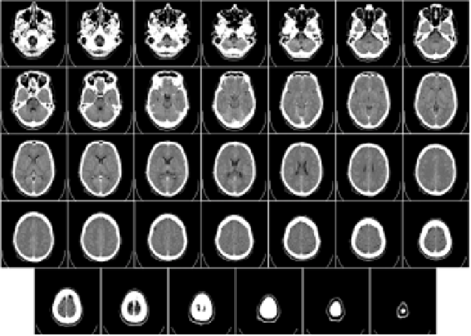
Fig. 12.2
Complete tomography image series of a brain
x-ray projections but in this case, the patient is enclosed in a surrounding ring of
detectors. Since its introduction in the 1970s, CT has become an important tool and
it is used to diagnose different diseases in the head (see Fig. 12.2), bones, lung, heart
vessels, etc.
Magnetic Resonance Imaging (MRI):
A magnetic resonance imaging instrument
(MRI scanner), uses powerful magnets to polarise and excite hydrogen nuclei (sin-
gle proton) in water molecules in human tissue, producing a detectable signal which
is spatially encoded, resulting in images of the body. The MRI machine emits a
radio frequency pulse that specifically binds only to hydrogen. MRI traditionally
creates a two dimensional image of a thin slice of the body. Modern MRI instru-
ments are capable of producing images in the form of 3D blocks, which may be
considered a generalisation of the single-slice, tomographic, concept. MRI is sensi-
tive to different tissue properties and has an excellent soft-tissue contrast. In clinical
practice, MRI is used to distinguish pathologic tissue (such as a brain tumour) from
normal tissue in every part of the body. MRI is an important research tool to map
normal human brain (see Fig. 12.3).
Nuclear Medicine:
Nuclear medicine encompasses both diagnostic imaging and
treatment of diseases, and may also be referred to as molecular medicine or molec-
ular imaging and therapeutics. Nuclear medicine uses certain properties of isotopes
and the energetic particles emitted from radioactive material to diagnose or treat var-
ious pathologies. Gamma cameras are usedine.g. scintigraphy,SPECTandPET