Biomedical Engineering Reference
In-Depth Information
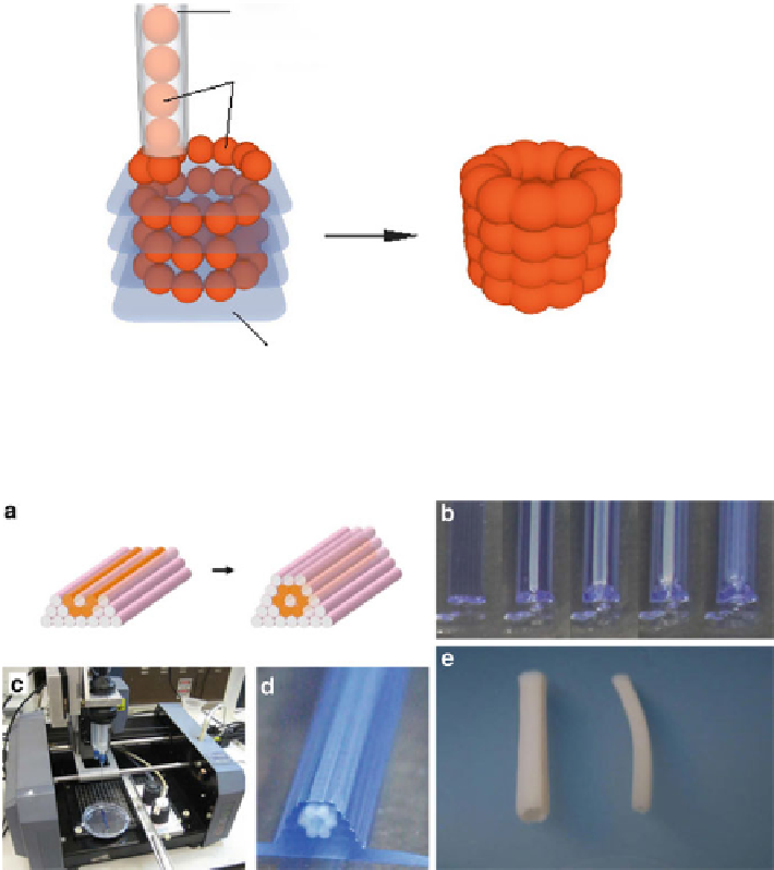
Nozzle
(xyz movement)
Ball of bio-ink
Bio-paper
Merging of the bio-ink particles
Fig. 9.10
Basic concept of bioprinting bio-ink particles into bio-paper (hydrogel) sheets. The bio-
ink particles are deposited in a tubular geometry (
left
). After the deposition is finished, the construct
is transferred to a bioreactor to fuse the bio-particles and further maturation made possible (
right
)
Fig. 9.11
Bioprinting tubular structures with cellular cylinders. (
a
) Designed print template
(
b
) Layer-by-layer deposition of agarose (
blue
) cylinders and multicellular pig SMC cylinders
(
white
). (
c
) The bio-printer outfitted with two vertically moving print heads. (
d
) The printed
construct. (
e
) Engineered pig SMC tubes of distinct diameters resulted after 3 days of post-printed
fusion (
left
: 2.5 mm OD;
right
: 1.5 mm OD). Pictures were reprinted from [
287
] with permission
from Elsevier
a poly[
N
-isopropylacryamide-co-2-(
N
,
N
-dimethylamino)-ethyl acrylate] copolymer
in a concentration of 10 wt.% polymer in cold, deionized water.
However, collagen used in a sheet-like design appeared to have integrated into
the final structure, posing difficulties in its removal [
291
]. Depending on the tar-
get tissue design, the bio-paper can also have other geometries. For instance, aga-
rose rods were plotted and easily removed after post-printing fusion of a
Search WWH ::

Custom Search