Biomedical Engineering Reference
In-Depth Information
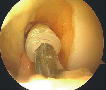
Fig. 6.4
Intraoperative—
arthroscopic image of the
interference screw during
insertion for anterior cruciate
ligament graft fixation in the
intercondylar notch
Fig. 6.5
Composite picture of a retrieved interference screw obtained from a series of SEM
images
Reamers that allow us to extract the PLLA screw without damage, as seen in Fig.
6.5
which presents a complex image of the screw using a mix of different SEM images
at the same scale.
SEM analysis suggested that the amount of threads engaging the bone plug dif-
fered in different screw designs. Figure
6.6
shows a progressive increase in cracks
and cavitations on the root and thread with time. Cracks appear first in the thread
with an axial orientation.
Also, SEM evaluation of the screw showed significant thread damage attribut-
able to bone plug pullout (Fig.
6.7
).
Another observation from the clinical practice was that the failure of the graft usually
occurs at the tunnel entrance site, therefore being possible for screw threads to produce
tissue laceration or lesions that cumulate during the postoperative rehabilitation.
Therefore, the SEM analysis of bioresorbable screws of PLLA showed that the
screw threads present no similarity to their original shape after retrieval (even if
degradation began for some time, the design should have similarities). Ultrastructural
analysis showed a gradual formation of cracks and cavities, over time, starting at the
top of the screw thread. It seems that the thread is due to external damage and
forcing the extraction.
In general, the degradation process includes progressive increase in cracks and cavi-
tation on the root and thread with time, which is consistent with anatomo-pathological

Search WWH ::

Custom Search