Biomedical Engineering Reference
In-Depth Information
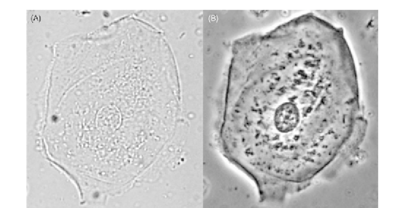
Figure 1.1
Comparison of a mammalian cell imaged with (A) brightfield and (B) phase contrast light
microscopy.
of microscopy. This chapter will explain how phase contrast works and how it is done
practically in standard microscopes; some typical uses of the technique are also presented.
1.2 How Phase Contrast Works
1.2.1 Basic Overview
Brightfield transmitted light microscopy has a fundamental weakness in the study of thin
transparent biological specimens: they generally absorb little light to provide limited
contrast by this means alone. The samples do however interact with light in ways other than
absorption, and phase contrast is a method to exploit this and turn these interactions into
observable contrast.
What does happen to light as it passes through transparent biological material such as the
cell in
Figure 1.1
? Though transparent, the cell will cause diffraction and scatter of some of
the light that passes through it. This process causes deviation and a phase shift of
/4
between the small amounts of light that is deviated relative to the undeviated wave. A cell
is far from homogeneous. The variety of components of the cell and the molecular,
macromolecular, and organelle level structures into which they are arranged provide a
complex landscape of changes in refractive index, and this variation will also have an effect
on the light passing through the different parts of the cell.
λ
Search WWH ::

Custom Search