Biomedical Engineering Reference
In-Depth Information
(A)
(B)
175
µ
m
175
µ
m
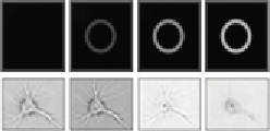
Figure 4.9
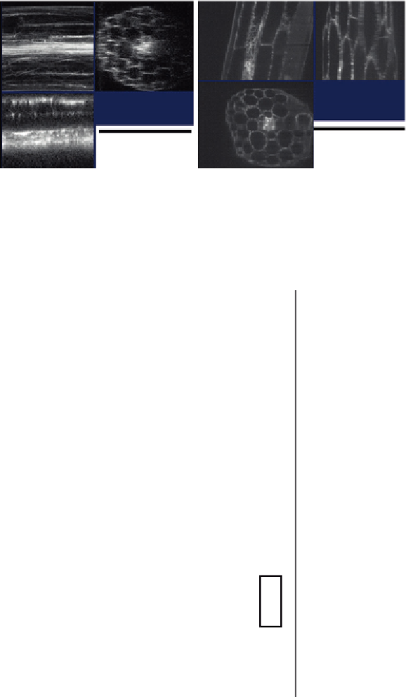
Adaptive optics in Arabidopsis root sections (A) as produced from fluorescence microscope
(B) after image processing.
Halogen lamp filament
Collector lens
Field
diaphragm
Aperture
Condenser
lens
SLIM module
Condenser
annulus
CCD
0
π
/2
π
3
π
/2
Specimen
Objective
Backfocal
plane
Fourier
lens L2
Phase rings
LCPM
Tube lens
Mirror
Image
plane
Beam
splitter
Fourier
lens L1
Figure 4.10
Configuration of SLIM microscope (left). The images of variation of phase rings as captured by
the CCD (right).


Search WWH ::

Custom Search