Biomedical Engineering Reference
In-Depth Information
(A)
(B)
Above
focus
Becke
line
Becke
line inside
Specimen
Below
focus
Becke
line outside
100
µ
m
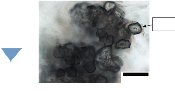
Figure 4.4
(A) Becke line principle (top) and its formation at the inside of specimen (bottom). (B) Image of
Becke lines in pear stone cells.
be formed inside the specimen at the above focus point. The formation of the Becke lines
inside the specimen is illustrated in
Figure 4.4A
.
A microscopical image that shows the formation of Becke lines inside the pear stone cells
is shown in
Figure 4.4B
. The stone cells are surrounded by the line in black around each
cell. This causes refractive index of cellulose encrusted with lignin and the depth of each
stone cell creating a large phase difference between the cell and its surroundings. The
surrounding medium is water which has a lower refractive index.
4.5 DIC Microscopy
Since the transparent specimen is not able to be visualized by the human eye, the
contrasting method plays an important role to overcome this problem. Of current
microscopical methodologies for optical microscopy, the DIC is the most widely used and
has fewest artifacts in imaging thick and absorbing specimens
[13]
. This kind of
microscope works by illuminating the transparent specimen with dual polarized light
sources that are split up from a single polarized light source via a Nomarski prism, such
that they become slightly incoherent (i.e., the offset is illustrated by the red and blue dash
lines that indicate the wavefront of each polarized light source) and perpendicular to each
other. The wavelength of the incident light changes as it passes through the transparent
specimen due to the refraction effect (usually becomes shorter due to the denser medium of
the transparent specimen), hence resulting in the changes of phase. While still not able to be


Search WWH ::

Custom Search