Biomedical Engineering Reference
In-Depth Information
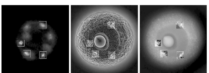
(A)
(B)
(C)
Figure 2.10
Simultaneous observation of live Spisula oocyte treated with DNA-specific fluorescent marker
(Hoechst 33342) by (A) fluorescence and (B and C) OI-DIC microscopy techniques—(B) gradient
and (C) phase (dry mass). Image size is 64
μ
m
64
μ
m.
3
chromosomes is somewhat larger than the fluorescence image because it shows both
proteins and DNA, while the fluorescence depicts stained DNA only.
This result indicates that OI-DIC can be used to visualize chromosomes in living cells and
provides sufficient detail to recognize chromosomes by their dynamic individual
three-dimensional shapes. OI-DIC imaging assessed the distribution of DNA in healthy
living cells without using fluorescent DNA markers and irradiating with excess excitation
light. Time-lapse imaging of DNA distribution during the mitotic phase of the cell cycle by
this noninvasive methodology is an important tool. Moreover, as OI-DIC provides a
two-dimensional distribution of the amount of DNA within an extremely thin optical
section, OI-DIC enables reconstruction of the three-dimensional distribution of DNA in
the nucleus.
Acknowledgment
This work was supported by NIH Grant Number R01-EB005710.
References
[1] F.H. Smith, Interference microscope, US Patent 2601175, (5 August 1947).
[2] F.H. Smith, Microscopic interferometry, Research (London) 8 (1955) 385.
[3] A.A. Lebedeff, Polarization interferometer and its applications, Rev. Opt. 9 (1930) 385.
[4] G. Nomarski, Interferential polarizing device for study of phase object, US Patent 2924142 (14 May 1952).
[5] R.D. Allen, G.B. David, G. Nomarski, The Zeiss
Nomarski differential equipment for transmitted light
microscopy, Zeitschrift f¨r Wissenschaftliche Mikroscopie und Mickroskopische Technik 69(4) (1969) 193.
Search WWH ::

Custom Search