Biomedical Engineering Reference
In-Depth Information
DIC4
Beam-shearing
assembly 2
LC3
DIC3
DIC2
LC2
Beam-shearing
assembly 1
LC1
DIC1
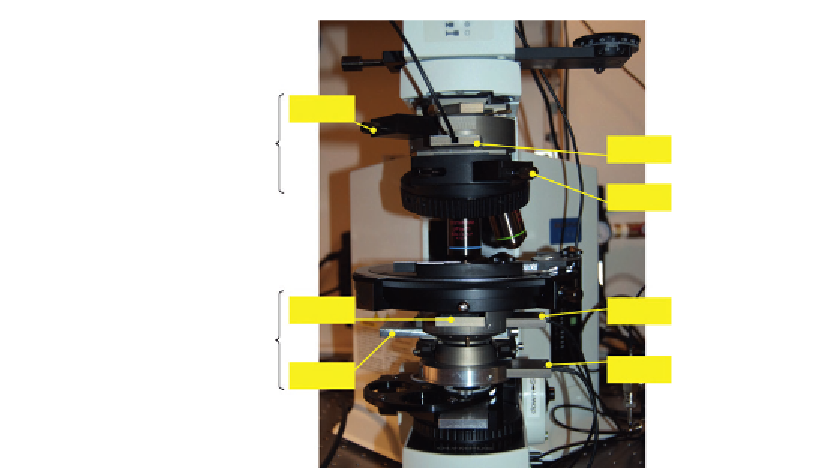
Figure 2.7
OI-DIC microscope setup with two switchable beam-shearing DIC assemblies. The first assembly
consists of DIC prisms DIC1 and DIC2 and two liquid crystal variable retarders LC1 and LC2. The
second beam-shearing DIC uses prisms DIC3 and DIC4 and liquid crystal variable retarders LC3.
Figure 2.8
Phase OI-DIC image of live crane fly spermatocyte at metaphase of meiosis I.
chromosomes are in sharp focus at the spindle equator, along with one of the
X
Y
sex
univalents, which is located on the right. The tubular distribution of mitochondria
surrounding the spindle is clearly evident. Both polar flagella in the lower centrosome are
in focus, appearing as a letter “L” lying on its side. The experiment was done together with

Search WWH ::

Custom Search