Biomedical Engineering Reference
In-Depth Information
18.4.1 Experimental Characterization of the Fluorescence Phase Microscopes
The Fluorescent Axial (Depth) PSF
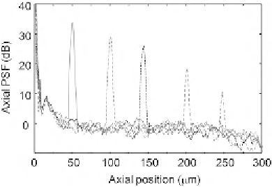
The full-width at half-maximum (FWHM) of the axial PSF of the FPM systems is an
adequate measure for the axial accuracy with which individual fluorophores or a dense
aggregate of fluorophores can be determined. The axial PSF FWHM can be obtained by
estimating the extent of the amplitude profile of the self-interference signal. To characterize
the axial PSF of the FPM systems, we used a single thin fluorescent layer made by drying
fluorescent nanoparticles on a coverslip and measured the spectrum or autocorrelation
function of the systems. Sharp axial PSFs with FWHM of approximately 3
m were
reconstructed by the SD-FPM setup shown in
Figure 18.3A
at shot-noise limited SNR
levels of 15
30 dB as shown in
Figure 18.4 [39,41]
. Similar FWHM values were also
obtained for the TD configuration
[39]
. A first-order approximation to the extent of a
Gaussian axial PSF is given by 2 ln 2
μ
2
0
are the peak wavelength
and bandwidth of the fluorescence emission spectrum, respectively. Under the Gaussian
PSF profile assumption, common fluorescent markers, such as cyan fluorescent protein,
4
0
,6-diamidino-2-phenylindole, and dimethylsulfoxide, would provide theoretical axial
resolution levels of 1.8 and 0.9
μ
m, respectively. We note, however, that non-Gaussian
profiles will result in the presence of sidelobes in the axial PSF that generate spurious
structures in the acquired images and mask weak fluorophores located near a strong
fluorophore. In addition, we point out that in contrast to the TD-FPM configuration, the
SD-FPM system resulted in a lower SNR level when the fluorescent layer was positioned
farther away from the zero differential optical path-length point of the interferometer (z
0
).
The reason for this effect is the finite resolution of the spectrometer that averages the
=πλ
=Δλ
where
λ
0
and
Δλ
Figure 18.4
Axial PSFs of a single layer of 100 nm fluorescent beads versus axial position of the layer. Sharp
axial PSFs with FWHM on the order of a few micrometers are clearly observed. Source: This
figure is reproduced from figure 4(a) of Ref.
[41]
with permission of the Optical Society of America.

Search WWH ::

Custom Search