Biomedical Engineering Reference
In-Depth Information
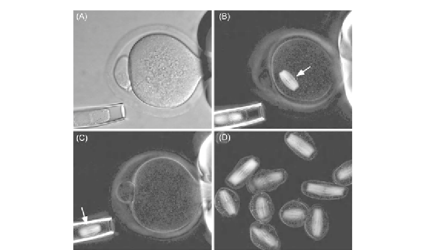
Figure 15.8
Mouse oocyte and enucleated spindles. (A) Mouse oocyte held in place by gentle suction of a
holding pipette (oocyte diameter approximately 75
m, DIC microscopy). (B) Retardance image
of the same oocyte, with birefringent spindle of meiosis II (white arrow). (C) The spindle
(arrow) is aspirated into an enucleation pipette. (D) A batch of enucleated spindles and
chromosomal karyoplasts. Chromosomes are aligned in the middle of spindles. Source: Courtesy
of Dr. Lin Liu
[42]
μ
Lenses and other optical elements that are part of the optical train but not located between the
crossed polarizers do not affect the polarization performance of the microscope setup. Filter
cubes for epi-illumination, for example, should be placed outside the polarization optical
train, if possible. (The polarization optical train is defined as the stretch between the crossed
polarizers.) In a dissecting microscope, the polarizing elements and compensator could be
placed in front of the objective lens instead of behind it. The objective of a dissecting scope
has very low NA and the image-forming rays have a small angle of divergence (in contrast to
high NA lenses). Rays with a small tilt angle with respect to the normal of the polarizer and
compensator plates do not appreciably affect the performance of these devices.
15.4.2 Specimen Preparation
The following are recommendations for preparing living cells for observation with a
polarizing microscope. Other resources for polarized light microscopy of living cells
include Refs.
[14]
and
[17]
, which have sections on suitable cell types and their preparation.
Search WWH ::

Custom Search