Biomedical Engineering Reference
In-Depth Information
6
5
4
3
2
1
0
-1
-2
-3
200
150
100
50
0
-50
-100
-150
-200
-250
-20
-10
0
10
20
30
40
50
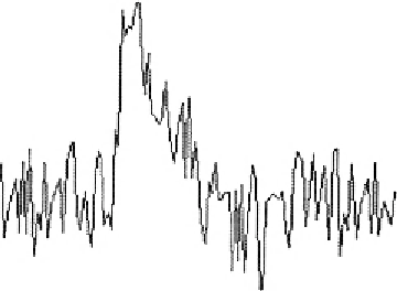
Time (ms)
Figure 13.7
Nerve displacement and electrical potential measured using the phase OCM system. Source: Taken
with permission from Ref.
[20]
. Copyright of Optical Society of America.
0.3 nm, at least three times better compared to the commonly used holographic phase
microscopy system under the same conditions. In single-point interferometric measurements
of live cells, the light is focused on a small area and therefore higher irradiance allows for
ultrafast acquisition times with compact and reasonable-price detectors. Furthermore,
single-point measurements also allow for an easy-to-implement common-path geometry and
a compact and portable fiber optics configuration, and thus can work even without using
floating optical tables, which has a significantly higher potential for clinical applications.
When measuring RBCs with quantitative phase microcopy, a constant RI assumption
can be taken, so that the phase measurement is proportional to the thickness of the RBC.
Using the time-dependent thickness profile of the RBC, cell stiffness properties can be
calculated.


Search WWH ::

Custom Search