Biomedical Engineering Reference
In-Depth Information
t
= 0 h
t
= 14.3 h
t
= 18.4 h
t
= 18.7 h
12 rad
Cell division
Cell division
t
= 32.5 h
t
= 33.5 h
t
= 32.4 h
t
= 19.7 h
0
Cell division
t
= 38.4 h
t
= 38.8 h
t
= 37.7 h
t
= 36.8 h
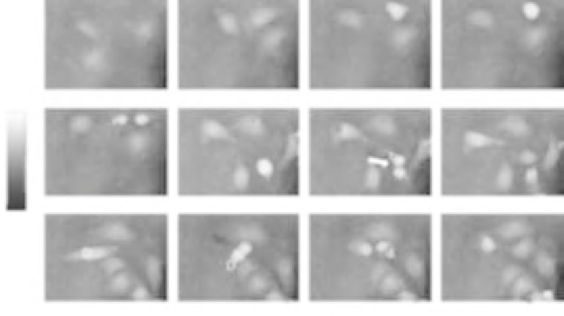
Figure 6.12
Time-dependent quantitative DHM phase contrast images of HBMECs (coded to 256 gray levels).
The arrows indicate cell division after t
37.7 h (cell
D). For cell C, no mitosis is observed during the experimental period. The corresponding daughter
cells after the cell division process are denoted as a
1
,a
2
,b
1
,b
2
,d
1
,d
2
, respectively
[82]
.
19.7 h (cell A), t
35.5 h (cell B), and t
5
5
5
particular for cell D at
t
5
37.7 h. A gray level coded pseudo-3D representation of the
quantitative contrast images of
Figure 6.12
is shown in
Figure 6.13
. For cell cycle phases in
which the cells adhere on the substrate, the quantitative phase contrast images correspond to
the cell shape (see
Eq. (6.4)
), while during cell rounding, the projection of the cell thickness
is measured (see
Section 6.4
and Ref.
[52]
).
For further evaluation of the DHM phase contrast images, the maximum phase contrast
Δϕ
cell,max
, the maximum cell thickness
d
cell,max
, and the cell migration trajectories of all
cells were determined. Therefore, in a first step, the phase distributions were low-pass-
filtered 2 times, with a box average filter of 5
3
5 pixels. In this way, substructures of the
specimen in the phase distributions and noise
e.g., due to parasitic interferences and
coherent noise
were reduced. Afterward, within a region of interest (ROI) in which the
analyzed cell was located, the pixel coordinates of the maximum phase contrast were
determined. The automated tracking of dynamic displacements of time-lapse sequences was
performed by successive recentering of the ROI to the coordinates of the preceding
maximal phase value
[83]
. During the evaluation procedure, the trajectories of the
remaining daughter cells that were not detected by the automated cell tracking algorithm
were started by manual selection after the cell division.
Search WWH ::

Custom Search