Biomedical Engineering Reference
In-Depth Information
the wave front of the object wave. The interferogram that is formed by the superposition of
object wave and reference wave is recorded by a charge-coupled device (CCD) camera and
transferred to an image processing system for reconstruction and evaluation of the digitized
holograms.
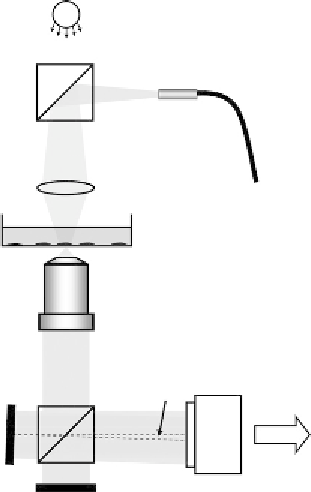
6.2.2 Self-Interference DHM
A drawback of many Mach
Zehnder-interferometer-based quantitative phase imaging
arrangements
[7,9
11,29]
is the requirement for a separate reference wave, which results in
a phase stability decrease and the demand for a precise adjustment of the intensity ratio
between object and reference waves. To overcome these problems, several approaches were
reported
[17,30
34]
. Here, in order to avoid a separately generated reference wave, a
Michelson interferometer approach for DHM is presented
[35]
.
Figure 6.2
shows a sketch of the Michelson interferometer-based DHM arrangement setup
which can be attached to an inverted research microscope
[35]
. In analogy to the setup in
WS
BS1
SM
CL
S
MO
α
BS2
PC
M2
M1
Figure 6.2
Schematic of an experimental setup for Michelson interferometer-based self-interference DHM.
WS: white light source; BS1, BS2: beam splitter cubes; CL: condenser lens; SM: single-mode fiber;
S: sample (here, Petri dish with adherent cells); MO: microscope lens; M1, M2: mirrors; CCD:
charge-coupled device sensor;
α
: tilt angle; PC: computer
[36]
.

Search WWH ::

Custom Search