Biology Reference
In-Depth Information
influence dendritic spine morphology and maintenance [Carlisle and Kennedy, 2005]. Taken
together, the observed disruption of dendritic spines links ADDLs to a major facet of AD
pathology, and provides compelling evidence that ADDLs in AD brain cause neuropil
damage believed to underlie dementia.
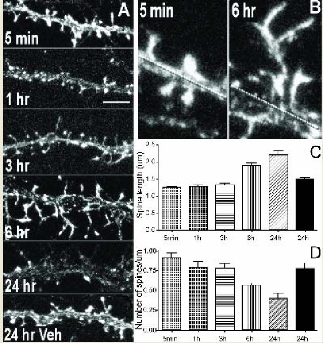
Figure 1. ADDL-induced aberrations in dendritic spine morphology and density. Cultured rat
hippocampal neurons at 21 days in vitro were treated for the times indicated with 500 nM ADDL.
A,
Confocal microscopy images representative of individual dendrtitic branches decorated with spiny
protrusions immunolabeled for drebrin after ADDL or vehicle (Veh) treatment. Longer and more
irregularly shaped spines appear after as early as 3 hours treatment and are more pronounced after 6
hours. Also note the reduced number of dendritic spines after 24 hours ADDL. Scale bar, 5 μm.
B,
Illustration of zoomed dendritic branches harboring “spines” demonstrates the pronounced lengthening
of dendritic protrusions after 6 hours of ADDL treatment. The line marks the dendritic shaft.
C,D,
Histograms represent average length and density of drebrin-labeled dendritic spines after ADDL
(patterned bars) or vehicle (black bars) incubation at various times. See Lacor
et al.
(2007) for further
details. [Reproduced from
The Journal of Neuroscience 27(4),
P.N. Lacor, M.C. Buniel, P.W. Furlow,
A.S. Clemente, P.T. Velasco, M. Wood, K.L. Viola and W.L. Klein, Aβ oligomer-induced aberrations
in synapse composition, shape and density provide a molecular basis for loss of connectivity in
Alzheimer's disease, 796-807 (Fig. 5), Copyright (2007), with permission from The Society for
Neuroscience].