Biology Reference
In-Depth Information
In 6-OHDA lesioned rats we observed a statistically significant increase in the diameter
of the synaptic ending in both axes; in the ipsilateral striatum this increase was evident since
the 3rd day after the lesion and reached to the maximum at day 30th (figure 10-A and B, and
figure 12-2). The contralateral striatum of 6-OHDA lesioned rats disclosed an increase in the
diameter at day 20-30 after the lesion; the increment persisted until the 120th day (figures 10-
A and B, and 11-3).
Postsynaptic target
As it is shown in figures 10-C and 11, the contralateral striatum presents similar values to
those found in the sham operated group, being the axospinous synaptic contact the most
common. In contrast, in the ipsilateral striatum, the axodendritic contacts prevailed.
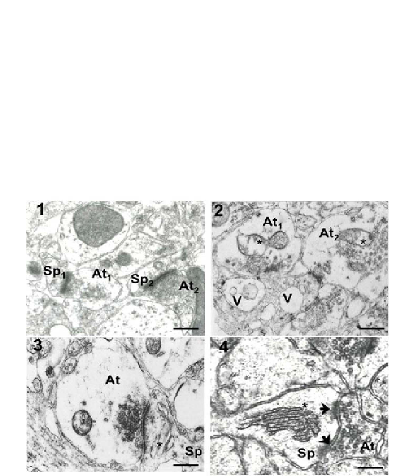
Figure 11. Electron micrographs from the striatum neuropil of the sham lesioned group (1); 6-ohda
lesioned ipsilateral striatum (2); 6-ohda contralateral striatum (3); 6-ohda lesioned ipsilateral striatum
(4). 1, In sham group, the mean size of the synaptic buttons (At
1
, At
2
) was 750 X 500 nm and the
predominant postsynaptic target was the dendritic spines (Sp
1
, Sp
2
), it can be observe that the neuropil
is well preserved 40,000 X. 2, This image corresponds to ipsilateral striatum neuropil and shows two
swollen synaptic buttons (At
1
, At
2
) and some vacuoles (V) within a dendrite; note the edematous
mitochondrion (*) 40,000 X. 3, This image demonstrates an edematous presynaptic ending (At) of the
contralateral striatum establishing a synaptic contact with a dendritic spine (Sp) with dilated spine
apparatus (*) 40,000 X. 4, An increase in the presence of perforated synaptic contacts was notorious in
both striata of lesioned animals (arrows). (At) Synaptic button; (Sp) dendritic spine; (*) spine apparatus.
40,000X. Bar: 0.25µm