Biology Reference
In-Depth Information
striatum contains both, projection neurons and several populations of interneurons (Bolam
and Bennett, 1995).
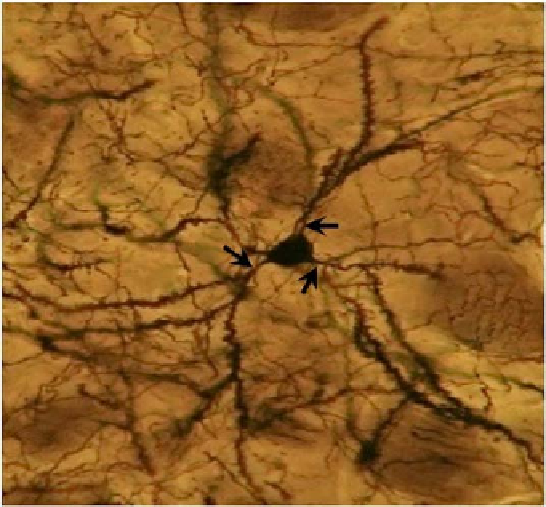
Figure 6. Light micrograph of a Golgi impregnated medium size spiny neuron in the rat striatum. Note
the medium sized perikaryon (approximately 20 µm in diameter), the spine-free proximal dendrites
(arrows) and the densely spiny secondary and higher order dendrites.
This neuron cell type, which constitutes over 90% of striatal neurons, is the major output
neuron of the striatum. Combined ultrastructural and neuroanatomical methods have
elucidated the organization of afferent connectivity to these neurons. The major physiologic
function of striatal efferent activity appears to be inhibition of tonically active GABAergic
neurons in the GP and SNr. Thus, the excitatory input from the cerebral cortex, whose
afferents make asymmetric synapses with the spines of MSN, appears to drive the efferent
activity of the striatum. Other extrinsic and intrinsic afferent synapses are situated in a
position to regulate the effect of the corticostriatal excitatory input to the MSN. For example,
dopaminergic afferents from the midbrain make mainly symmetric synapses with the spine
necks and dendritic shafts of the MSN. These neurons themselves have local axon collaterals,
which serve to link together local clusters of MSN. These local axon collaterals, which
contain, either GABA, substance P or enkephalin, also make mainly symmetric synapses with
the necks of spines or dendritic shafts of MSN. Other afferents with similar synaptic
connections to these neurons arise from cholinergic or somatostatinergic striatal interneurons.
Additionally, the patterns of extrinsic and intrinsic afferents to MSN and their extrinsic
projections are related to the organization of MSN into two mosaically organized
macroscopic compartments, the striatal patches and matrix (Gerfen, 1988). Figure 7
summarizes the synaptic organization of the medium spiny neuron.
Of particular importance, as we mentioned above, is the input from the dopaminergic
terminals derived from the SNc, which degenerate in PD. These terminals form symmetric