Biology Reference
In-Depth Information
account for the majority of postsynaptic sites in the vertebrate brain. Dendritic spines
typically are in excitatory synapses, and thus typically receive the neurotransmitter glutamate
from their partner axon. Spines are found on the dendrites of most principal neurons in the
brain, and are notably found in the pyramidal neurons of the cerebral cortex, the medium
spiny neurons of the striatum, and the Purkinje cells of the cerebellum.
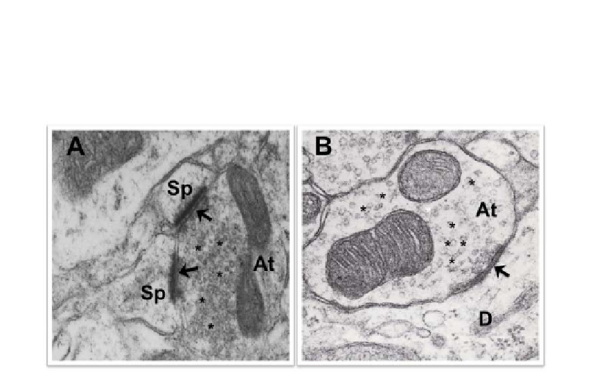
Figure 2.
A
. This picture shows an axon terminal (At) establishing an asymmetric synapse with two
dendritic spines (Sp). The synaptic bouton contains spherical vesicles (*). The arrows show the
postsynaptic electron densities.
B
. This electron micrograph shows an axon terminal (At) forming a
symmetric synapse with a dendrite (D). This synaptic bouton contains pleomorphic synaptic vesicles
(*). The arrow shows the postsynaptic electron density.
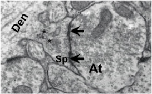
Figure 3. In this picture there is a spiny branchlet of a pyramidal cell dendrite (Den). The dendritic
spine (Sp) form asymmetric synapses (arrows) with the axon terminal (At). Spine can be recognized by
its spine apparatus (asterisks).
At ultrastructural level, spines can be distinguished from other dendritic elements in the
neuropil by the presence of a characteristic spine apparatus (figure 3) composed of a calcium-
binding protein, and it has been an important marker for spines in quantitative electron
microscopic studies. Within cerebral cortex, for example, about 79% of all excitatory
synapses are made onto spines and the rest directly onto dendrites, whereas 31% of all