Biology Reference
In-Depth Information
Also, there are examples of synapses that occur between two dendrites (dendro-
dendritic), between perikarya of two neurons (somato-somatic), and between perikarya and
dendrites (somato-dendritic and dendro-somatic).
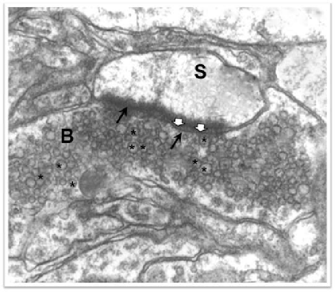
Figure 1. Electron micrograph of an axon terminal (B) that forms asymmetric synapse with a dendritic
spine (S). This axon terminal contains spherical vesicles (*). Note the membrane asymmetry (arrows)
called the postsynaptic electron density and the synaptic cleft (white arrowheads).
In the late 1950s Gray was the first to classify synapses on the basis of their junctional
characteristics. He referred to those synapses with prominent postsynaptic densities as type 1,
and described them as possessing a widened synaptic cleft, the separation between the faces
of the presynaptic and postsynaptic membranes being 20 nm wide (Fig. 2A); Gray also found
a different kind of synapse on the dendritic trunks and he referred to these as type 2 synapses.
Such synapses have a narrower synaptic cleft, about 12 nm wide, and dense regions of the
junction can be intermittent and have a less pronounced postsynaptic density than those of
type 1 synapses (Fig. 2B). In a later evaluation of the synapses came to conclusion that type 1
and type 2 synapses represent the extremes of a morphological continuum, and he chose to
refer them as asymmetric and symmetric synapses, on the basis of the disposition of the
cytoplasmic density on each side of the junction. Frequently, asymmetric synapses contain
round vesicles, and symmetric synapses contain both round and pleomorphic vesicles (Peters
et al., 1991).
The stereotypical and most abundant type of synapse in the central nervous system is the
asymmetric synapse occurring between an axon and a dendritic spine (Figure 2A).
The principal neurons of most brain regions are covered with small protrusions known as
dendritic spines. Spines are extremely numerous on many kinds of dendrites; in fact they