Biomedical Engineering Reference
In-Depth Information
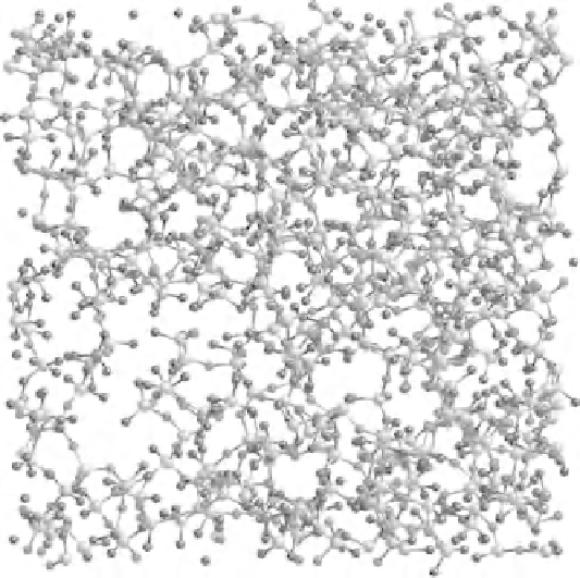
Figure 5.4
A snapshot of simulated 45S5 Bioglass, with the Na and Ca ions
removed. Although many of the PO
4
groups are not connected to the tetrahedral
network, there are some P-O-Si bonds. Si are pale gray/yellow, O are dark gray/red,
and P are larger and mid-gray/purple. For a better understanding of the figure, please
refer to the colour section (Figure 6).
information from scattering experiments. Recall that scattering data are
1D in nature, being a projection of the 3D structural information. It
is very hard, if not impossible, to recreate the 3D structure from the
1D scattering information. (Note that, if one is dealing with crystalline
materials, this is not the case, because the Rietveldt method of analyzing
powder diffraction data is available to one.)
The simulations support the idea that bioactive glasses have a highly
fragmented network structure, although the nature of the fragmentation
at NC
2 is more complex than simply chains of tetrahedra. Figure 5.5
shows the network fragments remaining after the largest fragment has
been removed from the structure in Figure 5.4. The simulations also
show that, as expected, in the most bioactive glasses there are a large
number of orthophosphate groups - those not bonded to any other
tetrahedra - and a significant number of orthosilicate groups as well.
However, not all of the PO
4
groups are found to be Q
0
species: some are
=