Biomedical Engineering Reference
In-Depth Information
50
45
HA R
2
=
0.99
40
35
BonAlive R
2
=
0.97
30
25
0
5
10
15
Time (months)
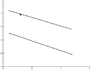
Figure 2.5
Resorption of BonAlive bioactive glass (BG) and hydroxyapatite (HA)
in vivo
. (Plotted from data in Ref. [19].)
reliable results, effective models are needed. The definitive animal model
for investigating the efficacy of bioceramics in bone regeneration was
developed by Oonishi
et al
. [18] using the rabbit femoral condyle (the leg
bone just above the knee). They compared the resorption of Bioglass to
that of synthetic HA and an apatite wollastonite glass-ceramic (AWGC)
in vivo
. After 12 weeks, 30-50% of the bioactive glass particles had
resorbed away and were shown to stimulate more bone growth than
AWGC and HA. Figure 2.5 shows the resorption of a bioactive glass
(BonAlive) and HA over a one-year period after implantation at two
different sites in rabbit frontal sinuses [19]. Approximately 30% of the
glass implants remained after 12 months (the difference replaced by new
bone). This is a good time frame for surgeons dealing with bone repair,
as it is important that the resorption of the implant does not happen
before the bone has the chance to regenerate.
In one of the only studies to compare the
in vitro
and
in vivo
behaviour
of bioactive glasses, Fujibayashi
et al
. [11] studied the bioactivity of a
series of ternary SiO
2
-Na
2
O-CaO glasses (i.e. phosphate-free). In this
case, the
in vitro
bioactivity (rate of HCA formation in SBF) decreased
as the silica content increased, which correlated well with
in vivo
response.
To investigate excretion of the dissolved silica from the body, Bioglass
excretion rates of silica were studied following implantation of Bioglass
in rabbit muscle [20]. The average excretion rate of silicon in urine
was 2.4mg/day (well below saturation). Excretion rates of silicon fell