Biomedical Engineering Reference
In-Depth Information
(a)
(b)
(c)
(d)
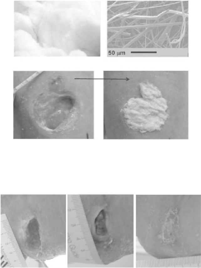
Figure 6.10
(a) A wad of bioactive borate glass nanofiber, (b) a high-magnification
SEM image of the fiber, (c) a wound prior to dressing with the borate glass nanofiber,
and (d) the same wound after dressing with the borate glass nanofiber. For a better
understanding of the figure, please refer to the colour section (Figure 7).
Figure 6.11
A chronic heel wound treated with borate glass nanofiber. Prior to
treatment, the wound had existed for two years.
The patient had the wound for over two years, was a diabetic, and in rela-
tively poor health. After approximately one month of treatments with the
borate glass fibers administered twice per week, the wound had resolved.
A second example is a venous stasis ulcer on the upper leg of an obese
woman in relatively poor health (prediabetic). The wound (Figure 6.12)
was initially presented with a strong odor (likely a yeast infection) and