Biomedical Engineering Reference
In-Depth Information
13-93B3
Front of skull
Suture
4mm
Implant
4mm
Implant
45S5
Sinus
Ear
Ear
Suture
Back of skull
(a)
(b)
Figure 6.7
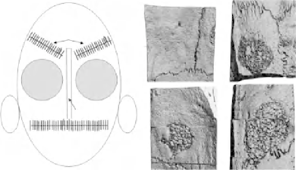
(a) Schematic of the rat skull showing the placement of the implants.
(b) Comparative X-ray microtomography images of 13-93B3 fiber scaffolds and
45S5 Bioglass particles after 12 weeks
in vivo
. The side of the CT image that is
labeled is the bottom side of the skull, and the unlabeled is the top of the skull. The
bottom side of the 13-93B3 scaffold is completely covered over with new bone and
cannot be seen. (Images acquired from Ref. [8].)
because silica-free bioactive borate glasses have been shown to stimulate
bone just as well as the silicate-based 45S5 Bioglass in rat calvaria
defects (Bi, L., Jung, S.B., Day, D.E.,
et al
., unpublished) [9]. Borate glass
(13-93B3) and 45S5 were compared in an identical bone growth model to
determine the effect that each has on bone regeneration. The model used
was the critical sized rat calvarial defect (4 mm), a schematic of which
is shown in Figure 6.7(a). After 12 weeks, X-ray microtomography was
used to visualize the defects, and a representative image from each glass
is shown in Figure 6.7(b). Although it is difficult to compare particles
with fibers, it was observed that the borate glass was completely covered
with new bone on the bottom side of the scaffold, whereas 45S5 Bioglass
was not.
Histological measurements of bone growth across the centre of the
implants (
N
4) are shown in Figure 6.8. The 45S5 and 13-93B3
are statistically similar, but the borate glass is completely devoid of
silica. The general consensus among those who study bioactive glasses
is that the presence of silica in bioactive glass is one of the most
important components for stimulating bone growth. The present data
are contradictory to the general consensus, in that a silica-free glass
was statistically just as beneficial
in vivo
as the well-known 45S5 glass.
=