Image Processing Reference
In-Depth Information
computer vision techniques. Forensic studies and biometrics (ways to recognise people)
using computer vision include automatic face recognition and recognising people by the
'texture' of their irises. These studies are paralleled by biologists and psychologists who
continue to study how our human vision system works, and how we see and recognise
objects (and people).
A selection of (computer) images is given in Figure
1.1
, these images comprise a set of
points or
picture elements
(usually concatenated to
pixels
) stored as an
array of numbers
in a
computer
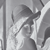
. To recognise faces, based on an image such as Figure
1.1
(a), we need to be
able to analyse constituent shapes, such as the shape of the nose, the eyes, and the eyebrows,
to make some measurements to describe, and then recognise, a face. (Figure
1.1
(a) is
perhaps one of the most famous images in image processing. It is called the Lena image,
and is derived from a picture of Lena Sjööblom in
Playboy
in 1972.) Figure
1.1
(b) is an
ultrasound image of the carotid artery (which is near the side of the neck and supplies
blood to the brain and the face), taken as a cross-section through it. The top region of the
image is near the skin; the bottom is inside the neck. The image arises from combinations
of the reflections of the ultrasound radiation by tissue. This image comes from a study
aimed to produce three-dimensional models of arteries, to aid vascular surgery. Note that
the image is very
noisy
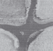
, and this obscures the shape of the (elliptical) artery. Remotely
sensed images are often analysed by their
texture
content. The perceived texture is different
between the road junction and the different types of foliage seen in Figure
1.1
(c). Finally,
Figure
1.1
(d) is a Magnetic Resonance Image (MRI) of a cross-section near the middle of
a human body. The chest is at the top of the image, and the lungs and blood vessels are the
dark areas, the internal organs and the fat appear grey. MRI images are in routine medical
use nowadays, owing to their ability to provide high quality images.
(a) Face from a camera
(b) Artery from ultrasound
(c) Ground by remote-sensing
(d) Body by magnetic
resonance
Figure 1.1
Real images from different sources
There are many different image sources. In medical studies, MRI is good for imaging
soft tissue, but does not reveal the bone structure (the spine cannot be seen in Figure
1.1
(d)); this can be achieved by using Computerised Tomography (CT) which is better at
imaging bone, as opposed to soft tissue. Remotely sensed images can be derived from
infrared (thermal) sensors or Synthetic-Aperture Radar, rather than by cameras, as in
Figure
1.1
(c). Spatial information can be provided by two-dimensional arrays of sensors,
including sonar arrays. There are perhaps more varieties of sources of spatial data in
medical studies than in any other area. But computer vision techniques are used to analyse
any form of data, not just the images from cameras.