Biology Reference
In-Depth Information
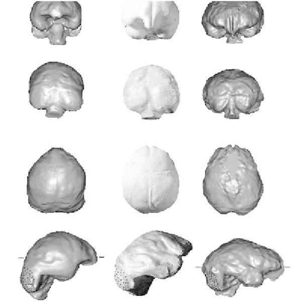
Figure 19. Endocasts, from left to right, of the microcephalic we described in
our
Science
report, the microcephalic(s) described by Weber's team, and LB1.
The endocasts are scaled to the same size for comparative purposes. Views:
(row
A) front, (row B) back, (row C) top, with frontal lobes oriented upward,
and (row D) right side, with frontal lobes oriented to the right. The speci-
mens have been correctly aligned, and the ones in the left and right columns
are virtual endocasts produced at Mallinckrodt Institute of Radiology. The
arrows in the last specimen in row A
point to the most anterior part of LB1's
BA 10. Other features include the transverse blood sinus
(t),
the sigmoid blood
sinus
(s),
frontal pole
(fp),
and occipital pole
(op).
The stippled areas in the
views in row D represent the cerebellum, which is tucked underneath the
occipital lobe and is located forward relative to
op
in LB1 but not in the two
microcephalics. Unlike nonpathological humans and LB1 (see figure 22), the
cerebellum is relatively large and protrudes posteriorly in the microcephalic
brains/endocasts. Image created by Kirk Smith, Mallinckrodt Institute of
Radiology; modified slightly from Falk, Hildebolt, et al., Response, 2005.