Agriculture Reference
In-Depth Information
abundant accumulation of secretion in the alveoli, indicating the onset of the lactogenic
process (Kensinger
et al.,
1982, 1986a). At the time of parturition, the lobules and alveoli
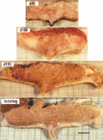
are completely filled with secretion (Figure 4.1; Turner, 1952). Figure 4.2 illustrates the
development of mammary tissue in pregnant gilts and a lactating sow. The location of the
gland on the udder affects its development during gestation. The wet weight of middle
glands (3
rd
, 4
th
and 5
th
pairs) is greater than that of posterior glands (6
th
, 7
th
and 8
th
pairs)
on both day 102 and day 112 of gestation (Ji
et al.
, 2006).
These phenotypic changes in the mammary tissue during late gestation coincide with
significant changes in mammary gene expression. In a study of the sow mammary
transcriptome, a number of pathways and gene networks were found to change through
the period between days 80 and 110 of gestation (Zhao
et al.
, 2013). For example, the
increased synthesis of milk lipid in mammary cells in late gestation may be driven by
activation of genes involved in fatty acid biosynthesis, the tricarboxylic acid cycle and
glyoxylate and decarboxylase flux. These analyses also indicate that there may be a
reduction in the degradation of essential amino acids and a reduction in other amino acid
metabolic pathways in late gestation, consistent with a dramatic increase in mammary
tissue protein deposition. Activation of genes associated with gap junctions, the mTOR
signaling pathway (milk protein synthesis), and VEGF and MAPK signaling (blood flow
regulation) all are consistent with known changes in mammary tissue function during
late gestation (Zhao
et al.
, 2013).
Figure 4.2. Transverse section of mammary glands from pregnant gilts during the last third of gestation and
lactation. Images represent days 80, 100 and 110 of gestation and day 3 of lactation. Images from gestation
are from the Hurley
et al.
(1991) study. Bar = 2 cm.