Geology Reference
In-Depth Information
by Ostwald's step rule (Jimenez-L
´
pez et al.
2001). According to this rule, stable phase for-
mation is sometimes preceded by metastable phases
which are normally favored under non-equilibrium
conditions (i.e. high supersaturation). Vaterite could
therefore be a precursor of calcite, forming in loca-
lized areas in which supersaturation and pH rapidly
rise as a consequence of local intense metabolic bac-
terial activity (Ben Chekroun et al. 2004). Local
increases in metabolic activity are consistent with
localized vaterite formation at high supersaturation
and also with the euhedral calcite rhombohedra
formed nearby at a lower supersaturation. A high
supersaturation seems to be a prerequisite for vater-
ite formation in the laboratory. This is consistent
with the observations of Rodriguez-Navarro et al.
(2003, 2007) regarding vaterite habit, size and
crystal density. The large number of tiny acicular
can be observed in the SEM photomicrographs
showing bacterial imprints within rhombohedral
calcite crystals (Fig. 4). In this study vaterite was
detected surrounding calcified bacterial cells
(Fig. 5). Vaterite is a metastable polymorph of
CaCO
3
and is rare in natural environments. It is
unstable and rapidly transforms into calcite (or ara-
gonite) at room temperature in an aqueous solution.
However, it commonly forms in synthetic processes
where organics are present and has been reported to
develop in the presence of microorganisms in nature
(see Rodriguez-Navarro et al. 2007, and references
therein). The presence of microbial films and the
apparent affinity of the bacterial cell walls for vater-
ite appear to cause vaterite precipitation in a manner
similar to that described by Mann et al. (1988).
One complementary explanation for vaterite devel-
opment in the presence of M. xanthus is suggested
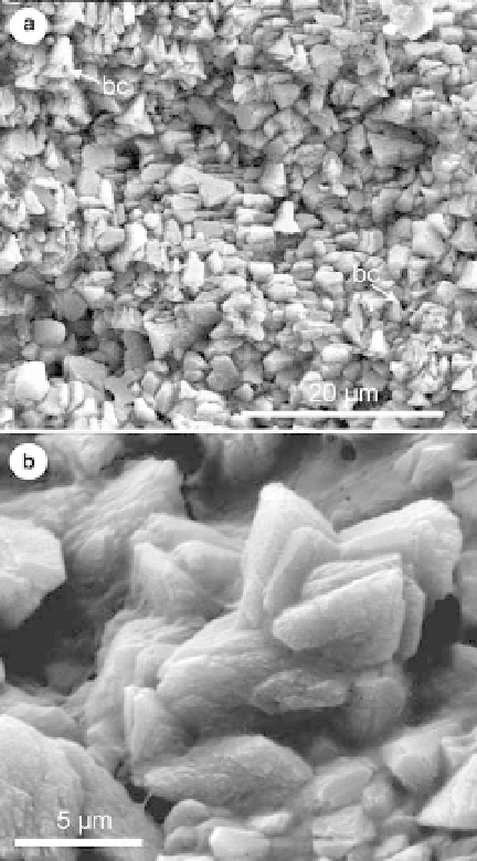
Fig. 4. SEM photomicrographs of: (a) calcite
rhombohedra with bacterial imprints (bc) developed on
calcarenite stone following a bioconsolidation treatment
with M. xanthus;(b) detail of bacterial
calcite rhombohedra.
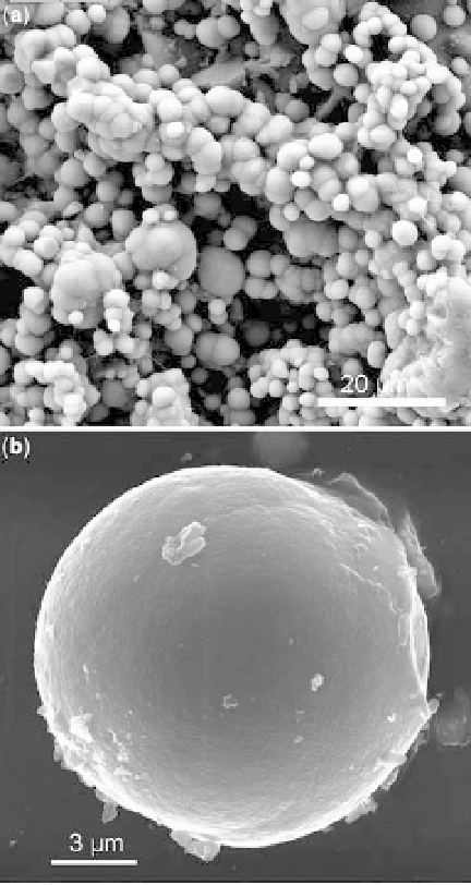
Fig. 5. SEM photomicrographs of: (a) aggregate of
vaterite spherulites formed in the presence of M. xanthus;
(b) detail of a bacterial vaterite spherulite.
Search WWH ::

Custom Search