Geology Reference
In-Depth Information
in Figure 1c and sub-parallel to the growth surface at
the top of the speleothem.
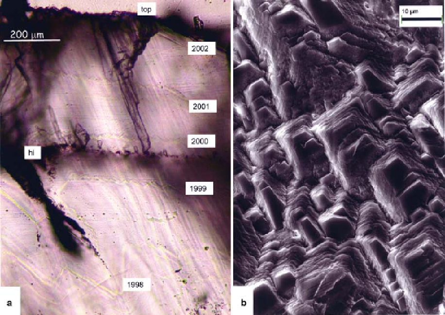
SEM analysis at the sample top (Fig. 3b) reveals
that the morphology of the calcite crystals is charac-
terized by sub-micrometre-scale macrosteps. Some
flat faces also developed which are apparently all par-
allel to each other. This contrasts with the more usual
situation in stalagmites (Frisia et al. 2000), where
SEM observations reveal that speleothem surfaces
are characterized by a variety of faces in different
orientations. The scaling indicates that each flat
face seen by SEM may correspond to a single crystal-
lite on the EBSD image. If the orientation analysed by
EBSD on a portion of the stalagmite holds true for
the rest of the specimen, as is supported by the ext-
inction patterns as seen by optical microscopy,
possible candidates for the flat faces which developed
at the top of the Obir stalagmites are those that lie
in the equatorial plane of the stereographic proje-
ction and pointing towards the top of the sample as
in Figure 2d. Plots for various prismatic, rhombohe-
dral and scalenohedral forms are shown and the
most likely
rhombohedral form (Fig. 2d; which differs from the
cleavage-parallel rhombohedron f10 - 14g). Later in
the paper, we interpret the presence of macrosteps
and the overall morphology of the calcite crystals
of Obir stalagmites as being controlled by the
presence of impurities in the parent solution.
Visible annual lamination
The top few centimetres of stalagmite Obi84 display
a consistent structure in which narrow visible
laminae, shown to be annual by Smith et al.
(2009), are spaced at c. 100 - 200 mm intervals.
Similar laminae are also developed in the other
two samples, but their development is less consist-
ent. The stalactite feeding Obi55 was collected
and also displays such laminae, but very close-
spaced. Where most distinctly developed in stalag-
mites, the laminae are a few microns wide and
may be nearly flat in geometry, but more commonly
display a zig-zag shape. Usually the zig-zags have a
relief of a few micrometres, but this can exception-
ally be up to 200 mm where it is followed by the
example
is
a face
in the f10 - 11g
Fig. 3. Petrology of sample Obi84. (a) ion probe thin section, transmitted light, illustrating a series of annual
event laminae with the last one being close to the top of the sample prior to collection in December 2002. Zig-zag
crystallite shapes are visible, and are of much higher relief in the 1998 layer, apparently corresponding to a pit on the
crystal surface which subsequently evolves into a (black) air inclusion. A within-year hiatus prior to the 2000 infiltration
lamina is marked by a re-nucleation horizon (hi). (b) SEM of top surface of sample illustrating crystallites which
display a mixture of flat smooth surfaces and rough surfaces representing stacked edges.
Search WWH ::

Custom Search