Geology Reference
In-Depth Information
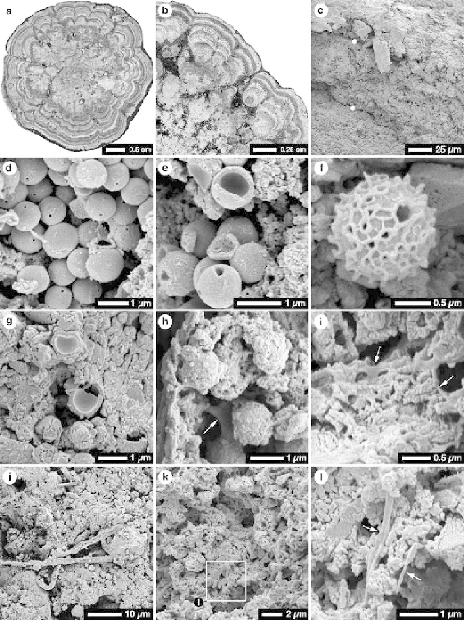
Fig. 8. Thin section (a & b) and SEM photomicrographs (c-l) of cave pisoliths from Old Man Village cave. (a, b) Thin
section photomicrograph of transverse sections through two cave pisoliths showing internal laminations. Note
'stromatolite-like' columns in outer part of pisoliths shown in Figure 8b. Dark layers formed of micrite, light layers
formed of spar calcite. (c) Peripheral micrite lamina (between white dots) in a pisolith that contains microbes and
textures shown in Figure 8d-l. (d) Group of smooth actinomycetes spores, each with a central open pore. (e) Smooth
actinomycetes amid micrite. Note thin wall of broken spore (upper part of image). (f ) Actinomycetes spore with
reticulate coating and pores. (g) Two broken actinomycetes spores buried in micrite groundmass. (h) Spherical bodies
Search WWH ::

Custom Search