Geology Reference
In-Depth Information
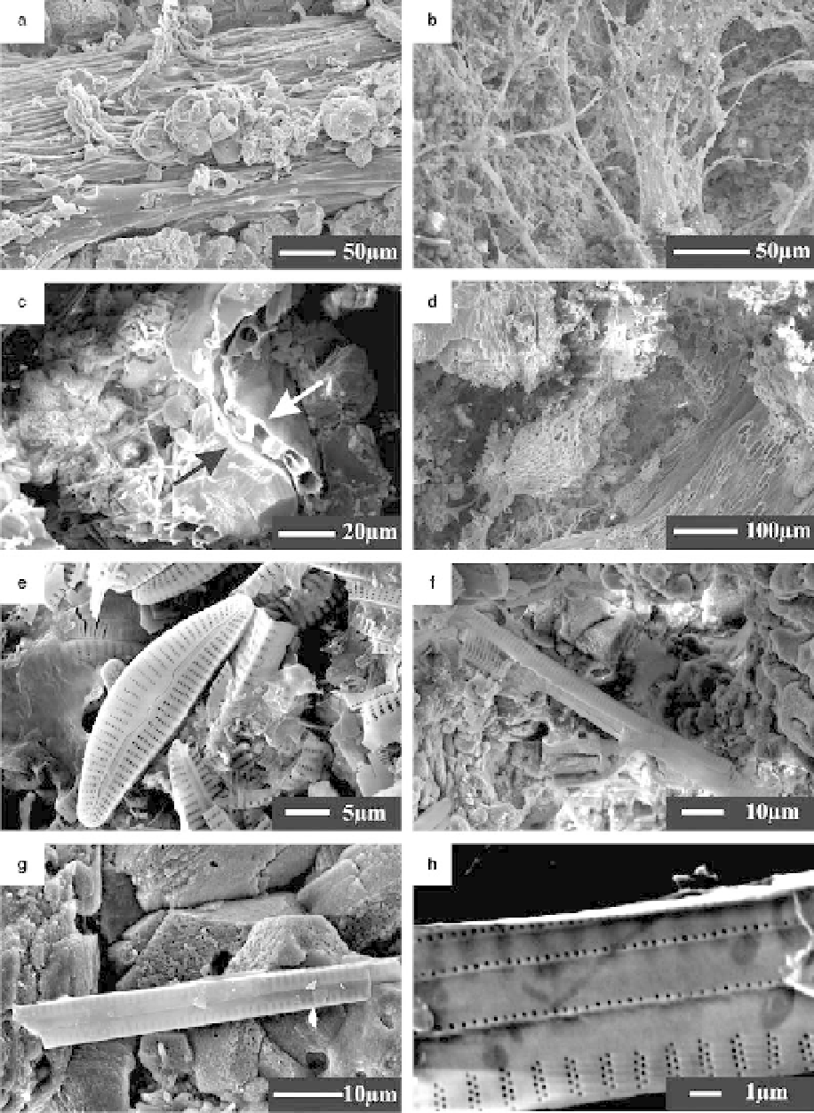
Fig. 8. SEM images of microbial components from the tufa deposits. (a) Longitudinal, fibrous microbial component
and adhered calcite crystals on its surface; (b) A network of microbial filaments and calcified extracellular polymeric
substances (EPS); (c) Curved leaf (between arrows) and diatoms in the middle; (d) Longitudinal cellular bryophyte
tubes; (e) Lenticular diatom frustules (Cymbella sp.), some diatoms deformed and broken; (f ) Rod-like diatom, Synedra
sp.; (g) Synedra sp. attached on calcite rhombs; (h) A close view of the diatom Synedra sp. in (g).
Search WWH ::

Custom Search