Geology Reference
In-Depth Information
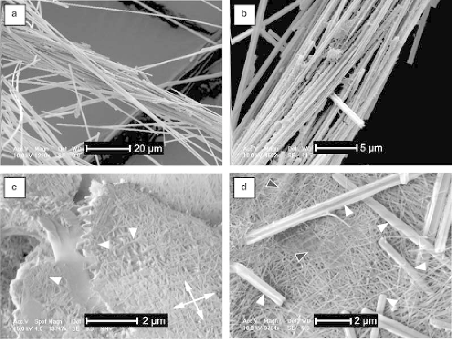
Fig. 3. (a-d) SEM photomicrographs. (a) Bundle of NFC present in a sample from the Spanish site of cotton-ball-like
NFC associated with a fungal strand (observed macroscopically). (b) Bundle of NFC covered by putative organic veils.
Some organic nanofibres are also present. Sample from a grain coating associated with a fungal strand and some
cotton-ball like NFC. (c) Close-up of an organized mesh composed by nanofibres (moonmilk, Swiss Alps). Preferential
orientations of nanofibres are shown by the crossed double-headed arrows. Some nanofibres are curved (arrows). This
characteristic indicates a contact-deformation. (d) Mesh composed by randomly-oriented nanofibres associated with
NFC (white arrows) and putative organic veils (black arrows). Swiss Jura Mountains, coatings on block.
often associated with other components (i.e. NFC,
fungal strands, hyphae, etc.) as observed in moon-
milk deposits (Fig. 3d); and (ii) organized structures
of nanofibres (Fig. 4), either as small pieces that
have apparently undergone a breakdown, or as a
tubular/circular microscopic network (Fig. 4a, c).
The term organized refers to a non-random distri-
bution of nanofibres, whatever their nature. These
networks are composed by intertwined nanofibres
oriented in two main directions (Fig. 4b, d).
Another main component is frequently observed
associated with soil samples: macroscopic, brown
organic filaments, identified as fungal strands.
Their average diameter can reach 100 mm and
they are composed of the two typical mycelial
strand structures, an external part made of several
narrow fungal hyphae with a thick cell wall and
an inner part characterized by a few wide thin-
walled hyphae, which often lack in our obser-
vations due to their ability to be rapidly decayed
(Fig. 5a, b). Nanofibres are abundant all along the
macroscopic filament where fungal strands seem
to break down.
A cross-section of a fungal hypha shows that the
fungal wall is composed of two layers, an inner part
composed of fibrous material and an external part
composed of an amorphous material (Fig. 5c, d).
From these observations, it is obvious that there is
an intimate relationship between the hyphae and
the nanofibres (Fig. 5c, d). Optical observations,
hydrochloric acid tests on moonmilk, as well as
TEM microdiffractions (Borsato et al. 2000) indi-
cate that the nanofibres are mineral in nature. In
order to test this hypothesis, in-situ analyses were
performed to distinguish organic from mineral
matter using osmium labelling with EDS control
on samples. The osmium stains only organic
matter and not mineral material (Pearson et al.
2004). Osmium peaks indicate that non-organized
frameworks composed of only nanofibres do not
Search WWH ::

Custom Search