Geology Reference
In-Depth Information
in some wood decaying species (Thompson 1984).
The structure of the fungal strand is composed
of: (i) an outer layer (the cortex) composed of a
thick layer of narrow thick-walled multiseptate
dead hyphae (average diameter of 1 mm); and
(ii) an inner layer (the medulla) composed of a
few linear wide thin-walled sparsely septate liv-
ing hyphae (average diameter of 6-10 mm). The
latter seems to be less resistant to hydrostatic
pressure (Watkinson 1979) and thus is more
rapidly exposed to decay processes than hyphae
with a thick melanized wall in the outer layer. The
inner part often collapses, leaving fungal strands
composed of a thick layer of narrow hyphae
with empty wide channels in the middle (Watkinson
1984). The cortex of the fungal strands makes it
an impermeable organ where fluids can be bidirec-
tionally translocated (Watkinson 1979; Dix &
Webster 1995).
et al. 1974; Farkas 1979; Ruiz-Herrera et al. 1996;
Pessoni et al. 2005). On the other hand, hyphae
from the cortex of a fungal strand can exhibit very
thick cell walls, up to 500 nm. The wall thickening
can even occlude the hypha lumen (Watkinson
1984). Moreover, the walls of these hyphae often
present a high degree of melanization, which
is also a factor in impermeabilization (Paul &
Clark 1996).
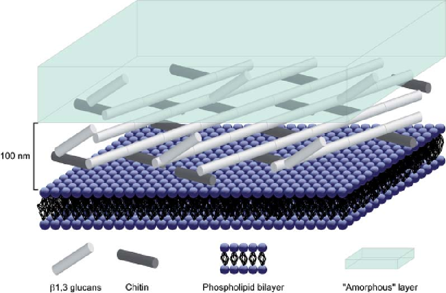
The fungal cell wall can be described by two
main types of materials, an outer layer composed
of amorphous material (mainly mannoproteins),
and an inner layer composed of fibrous material,
chitin and b-glucans (Burnett 1979; Ruiz-Herrera
1992; Bowman & Free 2006). Chitin is a polymer
of a polysaccharide, N-acetyl-glucosamine. It is
present in the form of long microfibrils, sometimes
over 1 mm, with a diameter of 10-25 nm. It is
located in the innermost part of the wall, arranged
as an intertwined mesh embedded in an amorphous
matrix (Aronson & Preston 1960; Carlile et al.
2001). b-glucans are homopolysaccharides of glu-
cose. In the fungal wall, it is present either as b
(1-3) glucan or in a lesser amount as b (1-6)
glucan. They are found in greater amounts than
chitin (Carlile et al. 2001; Farkas 2003). Figure 1
shows a sketch of the fungal cell wall.
Composition and structure of the
fungal cell wall
The thickness of the cell wall also shows great varia-
bility depending on physiological processes and the
function of hyphae. Single hyphae have an average
cell wall thickness of 150 nm (Jones 1970; Beckett
Fig. 1. Sketch of the fungal cell wall (modified from Latg´ 2003). Note the fibrous layer composed by chitin and
b-glucan fibres and the amorphous layer. In order to give an orientation to this sketch, the plasma membrane of the
fungal cell has been represented by the phospholipids bilayer.
Search WWH ::

Custom Search