Geology Reference
In-Depth Information
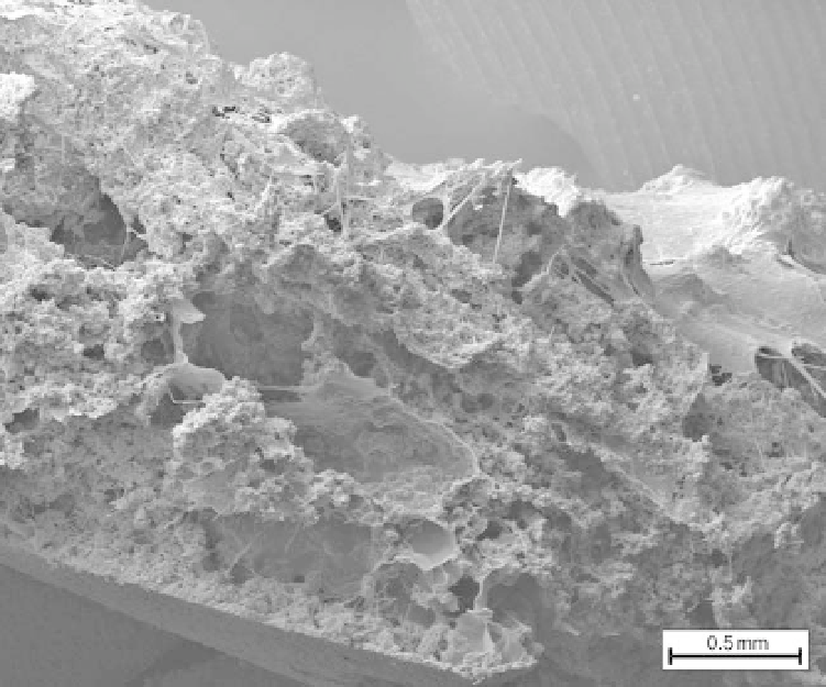
Fig. 9. Cross section of a laminated tufa biofilm grown under the uniform conditions throughout its 14 weeks
development (6 hours daylight and 10 8C temperature). Note the alternating layering between calcite dominated
(crystalline aggregates) and EPS dominated (dehydrated and cellular) areas. The biofilm shows a composite three layer
structure in which progressively younger basal calcite layers overbuilt the older cellular EPS fabrics to produce a
multilayered freshwater stromatolite. Autumn, fast-flow experiment. Air dried SEM sample.
described by Dupraz et al. (2004) in marine stroma-
tolites. Although the images presented by Dupraz
et al. (2004) showed these to be about 2 mm
wide (cf. 500 mm in the freshwater mesocosm
experiments) they share a similar style of calcite
nanospherulite development embedded within poly-
gonal EPS structures to that found in our exper-
iments. These polygonal structures appear too
similar to be coincidental and the difference in
scaling could be an important point to unravel, par-
ticularly if it is dependent on ionic diffusion gradi-
ents within the EPS which appears to be key to the
regulation of the water-biofilm-carbonate system
(Bissett et al. 2008).
Light microscopy of the living freshwater
biofilm revealed that the development of additional
layers (apparently achieved by calcite precipitation
within the outer surface of the biofilm) was inti-
mately related to the location of the tangle of dead
and coiled filamentous microbes. Precipitation in
the surficial part of the EPS has previously been
reported by Reid et al. (2000); Dupaz et al.
(2004), though from saline and hypersaline sites.
The driver for precipitation within the mesocosm
biofilms is unclear though it may also be triggered
by the EPS templating and dead cell degradation
by heterotrophic bacteria. Importantly, the multi-
layered mesocosm biofilm (Fig. 9) was seeded and
developed during a period of continuing fast-flow,
(autumn conditions) without any breaks to calcium
ion supply or in flow rate. This suggests that the
mesocosm freshwater stromatolite lamination was
controlled by organic factors internal to the
biofilm rather than by external seasonal variables
(see review in Andrews & Brasier 2005). Shiraishi
et al. (2008) has suggested that Ca
2þ
flux within
the EPS, caused by photosynthesis-induced calcite
precipitation coupled with microbial metabolic
functions, as the drivers for developing laminations.
Further experimentation is required before drawing
firm conclusions.
Growth of crystals within the mesocosm EPS
The microspar was predominantly developed from
nanospherulite precursors precipitated within the
EPS (cf. Fig. 3a). In few cases were nanospherulites
seen to be directly in contact with bacterial or
cyanobacterial sheath material nor was any replace-
ment of bacteria or cyanobacterial sheath observed.
Search WWH ::

Custom Search