Geology Reference
In-Depth Information
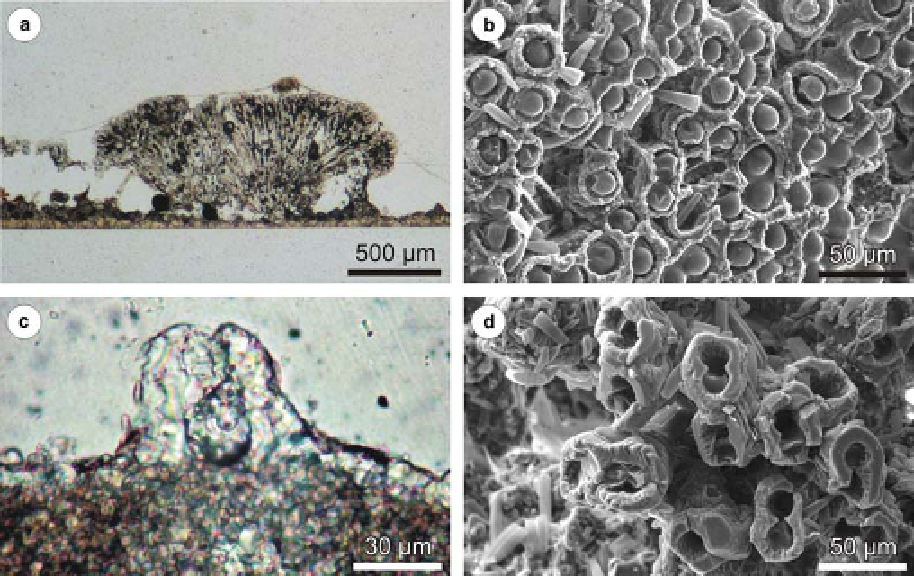
Fig. 14. Sparitic tubes of Oocardium stratum, all samples from the Z
´
zriv
´
site (a) bushy form comprising several
branching tubes that grew on Cu tablet exposed between August 2002 and October 2003, the tubes are divided from
Cu substratum by a sparry layer; (b) surface of Oocardium stratum colony seen from above, the cells are visible at the
tube tips, uncalcified diatom frustules occur on the sample surface, October 2003; (c) initial tube formed on limestone
tablet between August and October 2002; (d) twin tubes marking the cell division moment, surface of tufa, sample
collected in March 2003; a, c - thin sections, b, d - SEM image, lyophilized samples.
the Cayman Islands. However, they put forward
also other possible origins of micrite in a karst
environment.
filaments inside (Freytet & Verrecchia 1998; Pe-
ntecost & Whitton 2000). Also the general outline
of calcitized Phormidium colonies resembles the
described hemispheres (Pentecost 2003; Golubi´
et al. 2008). The coalescence and overlapping of
hemispheres
Hemispherically layered micrite. Micrite also forms
hemispheres up to 1 mm high and a few millimetres
wide (Fig. 19b, c). The hemispheres exhibit subtle
convex-up internal lamination visible most prob-
ably due to changes in microporosity. They com-
prise radially oriented cyanobacterial filaments
(Fig. 12d - f ). Minute calcite crystals are present
between the filaments. Detrital grains, including
quartz, are trapped there and between the hemi-
spheres (Figs 12f & 19d). The hemispheres occur
mainly at both the studied points of the Karw ´w
site. They occur in patches, but tend to be concen-
trated along the tablet edges (Fig. 19c, e, f ). They
predominate on the air-facing side of tablets but
some are also located on the substrate-facing one.
On the tablet exposed throughout the whole period
of the experiment the neighbouring hemispheres
coalesce into a continuous layer (Fig. 19e, f ).
The micrite hemispheres are akin to tuft-shaped
colonies of Phormidium incrustatum. What they
have in common is the size, internal lamination
and the presence of radially arranged cyanobacterial
result
from
horizontal
invasion
of
cyanobacteria (Pitois et al. 2001).
Fibrous micrite/sparite. Crystals from a few micro-
metres to .100 mm in size, encrusting algal fila-
ments, build porous tufa of fibrous texture
(Fig. 20a, b). The texture may be visible with the
naked eye (Fig. 20c). The filaments acting as a sub-
strate for nucleation belong to Vaucheria and some
cyanobacteria. Diatom frustules occurring within
this type of tufa become less numerous downward
the samples, probably because of dissolution. The
filaments are preserved as empty tubes.
Crystal size in filaments tends to increase out-
wards (Fig. 20d). Though intercrystalline porosity
is an important characteristic of this texture,
sizes of pores are variable. They depend on the
degree of cementation and on the primary arrange-
ment of the filaments (Golubi´ et al. 2008). If the
filaments are tightly packed, a small amount of
sparry calcite can effectively cement the whole
Search WWH ::

Custom Search