Geology Reference
In-Depth Information
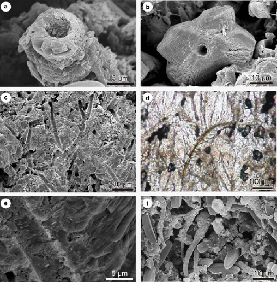
Fig. 11. Calcified cyanobacterial filaments: (a) micrite encrusted filament, Karw´w cascade point, December 2002; (b)
sparite encrusted filament, L ´ˇky top point, tufa formed on limestone tablet exposed between August 2002 and October
2003; (c) longitudinal section of filaments, L´ ˇky E point, tufa formed on Cu tablet exposed between August 2002 and
October 2003; (d, e) filaments serving as nucleation surfaces for calcite crystals, d - L´ ˇky E point, tufa formed on
limestone tablet exposed between August 2002 and October 2003, e - L´ˇky E point, tufa formed on Cu tablet exposed
between August 2002 and October 2003; (f ) parallely arranged living filamentous cyanobacteria partly covered with
calcite crystals, L´ ˇky E point, March 2003; all but d SEM images, d - thin section, f - lyophilized sample.
tablets at the H
´
j lower waterfall point and at the
Karw
´
w cascade point.
(Fig. 17a - c). The voids were detected at the H
´
j
lower waterfall, H
´
j dam points and at the L ´
ˇ
ky
site. They may be arranged in horizons within
the tufa samples as was the case at the H
´
j site or dis-
tributed randomly within the tufa section (Fig. 17b,
c). Some voids are less regular; they were probably
corroded (see Golubi
´
1969).
The voids are comparable to the larval housings
described
Larval housings. Vaulted voids up to 0.5 mm wide
and 0.35 mm high occur within the tufa deposited
on some tablets (Fig. 17). The voids have circular
or semicircular cross-section with convex-up
ceilings. They are covered with a thin lamina of
micrite, which in turn is overlain by sparry calcite
crystals
from many
tufa sites
in
Europe,
for
displaying
competitive
growth
pattern
instance
in
Germany
(Wallner
1934a;
Irion
&
Search WWH ::

Custom Search