Geology Reference
In-Depth Information
of Oocardium stratum cells (Fig. 14d; Golubic
et al. 1993).
Diatoms. The diatom flora is abundant in the studied
sites (T. Mrozi ´ska, pers. comm., 2003). It is docu-
mented by SEM observations of growing tufa
surface (Fig. 15a, b). The diatoms are associated
there with copious amounts of EPS (extracellular
polymeric secretions), some filamentous cyanobac-
teria and other algae. The diatoms belong to differ-
ent
morphological
groups.
Particularly
common
are
pennate
diatoms,
some
of
which
produce
slimy stalks.
The diatom frustules are discernible also within
the deposited tufa. They are particularly common
in micrite tufa, but have been also found in sparry
crystals (Fig. 15c). They are engulfed by calcite
but seldom act as a substrate for nucleation of
calcite crystals (Fig. 15d). Diatom frustules
without calcite nucleated on them was reported
from many modern tufa sites, and it seems to be
a rule (e.g. Merz-Preiss & Riding 1999; Lu
et al. 2000). In the course of the present study,
nucleation of crystals on diatom frustules was
observed exclusively in some samples from the
L ´ˇky site. In the deeper zones of the studied tufa
samples, frustules become less abundant, though
their moulds are found under the SEM (Fig. 15e;
cf. Pedley 1994; Szulc & Smyk 1994). It implies
that silica frustules can be dissolved within three
months or less.
Other microalgae. In the tufa sample from the
L ´ˇky E point, single calcified ovoid bodies 20 -
25 mm across were found. Inside, each of them con-
tained several small globules, 1 - 4 mm in diameter
(Fig. 15f ). The bodies strongly resemble colonies
of unicellular cyanobacteria that are common in
tufa-forming environment. At L ´ˇky they were
found in a high-energy setting in the waterfall
wall, in a shelter cavity formed by a leaf projecting
from the tablet and cemented to it. It seems probable
that they were earlier transported by the stream.
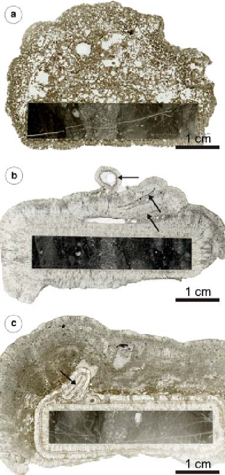
Fig. 9. Scanned thin sections of tufa formed on tablets,
(a) Karw´ w cascade point, tablet exposed between
August 2002 and October 2003, tufa displays high
porosity; (b)L´ ˇky E point, tablet exposed between June
2003 and October 2003; (c)L ´ˇky E point, tablet
exposed between August 2002 and October 2003; arrows
in b and c indicate leaves incorporated into tufa and
acting as additional nucleation surface.
Extracellular polymeric secretions. The extracellu-
lar polymeric secretions (EPS) cover the surface of
all samples. Under SEM they are visible as irregu-
larly twisted filaments or a three-dimensional reticu-
late structure (Fig. 16a, b; cf. D´farge et al. 1996).
The filaments are between ,1 mm and c. 15 mm
across. In some cases EPS construct a dense layer
which completely obliterates their substrate. The
EPS are spatially related to diatoms and filamentous
cyanobacteria. It also suggests a genetic relation-
ship, because some diatoms and cyanobacteria are
known to be efficient EPS producers (Riding 2000).
growth mode of its calcified tubes. After World
War II many other recent tufa sites abounding
in Oocardium stratum were recognized in Croatia,
France, Switzerland, Belgium, Poland, Great
Britain, USA and China (Golubi
´
& Mar
ˇ
enko
1958; Pentecost 1990, 1991; Freytet & Plet
1991; Mrozi
´
ska 1992; Golubic et al. 1993;
Pentecost & Zhang 2000; Golubi
´
et al. 2008).
The observed twin-tubes are a record of divisions
Moss stems. Moss stems were occasionally found
within the tufa deposited on the tablets. The moss
Search WWH ::

Custom Search