Image Processing Reference
In-Depth Information
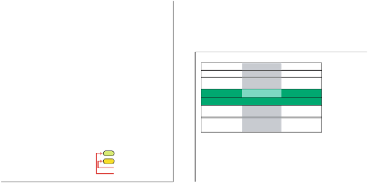
K6
K5
K4
R
6
L
5
R
4
L
3
Right
Left
Right
2
L
1
2
3
4A
4B
5
6
1
R
K3
K2
K1
Primary visual cortex (V1)
L: Left eye
R: Right eye
K1-6:
Interlaminar
zones
Parvocellular-Left
Parvocellular-Right
Magnocelular-Left
Magnocelular-Right
Konicelular-Left
Konicelular-Right
Parvocellular layers
Magnocellular layers
Ganglion (parasol) cells
Ganglion (midget) cells
LGN
Fig. 1.4. The
left
graph illustrates the left LGN of the macaque monkey with its six layers.
The
right
graph shows the left V1 and some of its connections, following Hassler labelling of
the layers [47, 109].
1.6 The Primary Visual Cortex
Outputs from each of the three LGN neuron types feed via optic radiations into dif-
ferent layers of the
primary visual cortex
, also known as
V1
,or
striate cortex
.TheV1
area has six layers totalling
2mmonafewcm
2
. It contains the impressive
200
million cells. To compare its enormous packing density, we recall that the ganglion
cells total
≈
≈
1 million in an eye. The V1 area is by far the most complex area of the
brain, as regards layering of the cells and the richness of cell types.
A schematic illustration of its input-output connections is shown in Fig. 1.4 us-
ing Hassler notation [47]. Most of the outputs from magnocellular and parvocellular
layers of the LGN arrive at layer 4, but to different sublayers, 4A and 4B, respec-
tively. The cells in layer 4A and 4B have primarily receptive field properties that are
similar to magnocellular and parvocellular neurons, which feed into the former. The
receptive field properties of other cells will be discussed in Sect. 1.7. The koniocellu-
lar cell outputs feed narrow volumes of cells spanning layers 1-3, called
blobs
[155].
The blobs contain cells having the so-called
double-opponent color property
. These
are embedded in a center-surround receptive field that is presumably responsible
for color perception, which operates fairly autonomously in relation to V1. We will
present this property in further detail in Sect. 2.3. Within V1, cells in layer 4 provide
inputs to layers 2 and 3, whereas cells in layers 2 and 3 project to layers 5 and 6.
Layers 2 and 3 also provide inputs to adjacent cortical areas. Cells in layer 5 pro-
vide inputs to adjacent cortical areas as well as nonadjacent areas, e.g., the superior
colliculus. Cells in layer 6 provide feedback to the LGN.
As to be expected from the compelling evidence coming from photoreceptor,
ganglion, and LGN cell topographic organizations, the visual system devotes the
largest amount of cortical cells to fovea even cortically. This is brilliant in the face
≈