Image Processing Reference
In-Depth Information
Time
Rod
Cone
Horizontal cell
Bipolar cell
Amacrine cell
Ganglion cell
Stimulus
Optic nerve
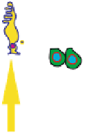
Fig. 1.3. The graph on left illustrates the retinal cells involved in imaging and visual signal
processing. On the right the response pattern of a (+
/−
)-type ganglion cell is shown
to the brain. All further processing in the brain takes place on “differential signals”,
representing local comparisons within and between the photoreceptor responses, not
on the intensity signals themselves.
The outputs of the ganglion cells converge to eventually form the
optic nerve
that goes away from the eye. Because the ganglion layer is deep inside the eye and
farthest away from the eye wall, the outputs come out of the eye through a “hole”
in the retina that is well outside of the fovea. There are no photoreceptors there.
The visual field region that projects on this hole is commonly known as the
blind
spot
. The hole itself is called the
optic disc
and is about 2 mm in diameter. Humans
actually do not see anyting at the blind spot, which is in the temporal hemifield, at
approximately 20
◦
elevation close to the horizontal meridian.
Exercise 1.1.
Close your left eye, and with your right eye look at a spot far away,
preferably at a bright spot on a dark background. Hold your finger between the
spot and the eye with your arm stretched. Move your finger out slowly in a half
circle without changing your gaze fixation on the spot. Do you experience that your
finger disappears and reappears? If so, explain why, and note at approximately what
elevation angle this happens. If not, retry when you are relaxed, because chances are
high that you will experience this phenomenon.
The ganglion cells are the only output cells of the eye reaching the rest of the
brain. There is a sizable number of retinal ganglion cell types [164], presumably to