Biomedical Engineering Reference
In-Depth Information
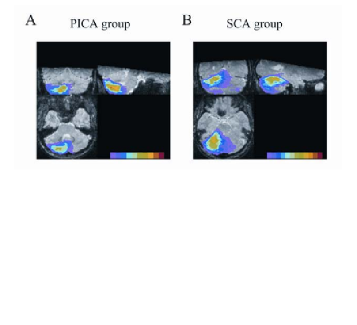
Figure 2
. Lesions of PICA (
A
) and SCA (
B
) patients superimposed on axial, sagittal, and
coronal stereotaxically normalized MR images of the cerebellum of a healthy 26-year-old
female subject. All unilateral lesions are superimposed on the left cerebellum, with right-sided
lesions flipped to the left. The number of overlapping lesions is illustrated in color. PICA
group: from violet (
n
= 1) to red (
n
= 11). Data of two patients are not included for technical
reasons. Note the center of overlap (orange,
n
= 10) in the posterior inferior cerebellum within
lobules VIIB and VIIIA. Note that some lesions affected the lower and inferior part of the
dentate nucleus (dark blue,
n
= 4; light blue,
n
= 5), with the interposed nucleus being pre-
served. SCA group: from violet (
n
= 1) to red (
n
= 12). Data from two patients are not included
for technical reasons. Note the center of overlap (light green,
n
= 9; darker greens,
n
= 8 and 7)
in the superior cerebellum within hemispheric lobules VI and Crus I.
involved in classical eyeblink conditioning. Eyeblink conditioning has been
shown to be impaired in patients with cerebellar lesions (10,17,75,80). In addi-
tion, studies using positron emission tomography (PET) and functional magnetic
resonance imaging (fMRI) revealed learning-related changes of activity in the
cerebellum during eyeblink conditioning in healthy human subjects (e.g.
(63,58)).
A recent human lesion study conducted by our group investigated classical
delay eyeblink conditioning in 27 patients with primarily unilateral lesions, par-
ticularly infarcts of the superior cerebellar artery (SCA) and the posterior infe-
rior cerebellar artery (PICA) (22). The extent of the cortical lesion (i.e., which
lobules were affected) and possible involvement of the cerebellar nuclei was
determined by 3D-magnetic resonance (MR) imaging. Figure 2B shows the le-
sions of all SCA patients and Figure 2A of all PICA patients superimposed on
MR images of the cerebellum of a healthy subject.
The cerebellar areas known to be most critical in eyeblink conditioning
based on animal data (i.e., Larsell lobule H VI and interposed nuclei) are com-
monly supplied by the superior cerebellar artery (4). Therefore, we hypothesized
that conditioning of the eyeblink reflex was impaired in patients with lesions