Biomedical Engineering Reference
In-Depth Information
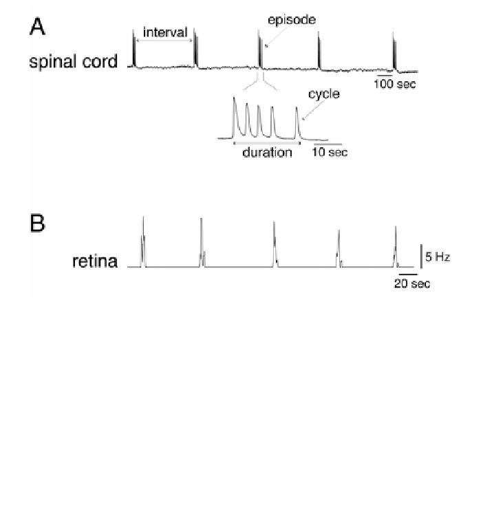
Figure 1
. Episodic activity in developing networks. A, Spontaneous activity recorded from the
isolated spinal cord of a 7.5-day-old chick embryo. The activity is recorded from a ventral root
and represents the synchronous activation of a population of motoneurons. It is characterized
by rhythmic episodes lasting up to a minute that are separated by silent intervals lasting up to
20 minutes. The high-frequency (fast) signal corresponding to motoneuron discharge is not
visible because of the scale and the low sampling rate (20 Hz). Modified with permission from
Tabak et al. (36). (
B
) Spontaneous activity recorded from the isolated retina of a 9-day-old
mouse (postnatal). This signal is the rate of neuronal discharge averaged over 29 cells. Note the
difference in the time scale with the recording in A. Data reprinted from J. Demas, S.J. Eglen
and R.O.L. Wong (unpublished).
vidual neurons (15,22). It is therefore important to understand the mechanisms
of generation of spontaneous activity in the developing nervous system. In the
following, we focus on the temporal organization of activity through activity-
dependent synaptic depression in the developing spinal cord, but suggest that the
same features are common to other neural networks.
Spontaneous activity was first observed as spontaneous movements in em-
bryos of diverse animals. Embryonic motility was extensively studied in the
chicken embryo (16), as it was easy to observe spontaneous movements through
the egg shell, and later to record electrical activity through a small window in
the shell. It was shown that these episodic movements were caused by spontane-
ous electrical activity in the neuronal networks of the spinal cord. More recently,
an isolated in vitro preparation of the embryonic chick spinal cord was devel-
oped (26), allowing one to record the activity and to manipulate the network (by
lesions or pharmacology) at different stages of development.