Biomedical Engineering Reference
In-Depth Information
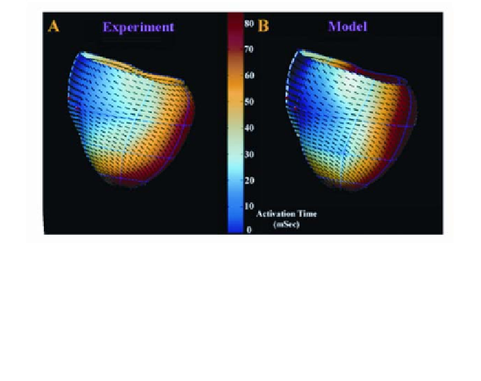
Figure 5
: (
A
) Electrical activation times (indicated by color bar) in response to right RV pac-
ing as recorded using electrode arrays. Data was obtained from a normal canine heart that was
subsequently reconstructed using DTMRI. Activation times are displayed on the epicardial
surface of a finite-element model fit to the DTMRI reconstruction data. Fiber orientation on the
epicardial surface, as fit to the DTMRI data by the FEM model, is shown by the short line
segments. (
B
) Activation times predicted using a computational model of the heart mapped
in
A
.
Use of DTMRI for reconstruction of cardiac fiber orientation provides sev-
eral advantages over traditional histological methods. First, DTMRI yields esti-
mates of the absolute orientation of cardiac fibers, whereas histological methods
yield estimates of only fiber inclination angle. Second, DTMRI performed using
formalin-fixed tissue: (a) yields high-resolution images of the cardiac bounda-
ries, thus enabling precise reconstruction of ventricular geometry using image
segmentation software; and (b) eliminates flow artifacts present in perfused
heart, enabling longer imaging times, increased signal-to-noise ratio, and im-
proved spatial resolution. Third, DTMRI provides estimates of fiber orientation
at more than one order of magnitude more points than is possible with histologi-
cal methods. Fourth, reconstruction time is greatly reduced (~60 hours versus
weeks to months) relative to that for histological methods.
DTMRI data acquisition and analysis for ventricular reconstruction has
been semi-automated. Once image data are acquired, software written in the
MatLab programming language is used to estimate epicardial and endocardial
boundaries in each short-axis section of the image volume using either the
method of region growing or the method of parametric active contours (65).
Diffusion tensor eigenvalues and eigenvectors are computed from the DTMRI
data sets at those image voxels corresponding to myocardial points, and fiber
orientation at each image voxel is computed as the primary eigenvector of the
diffusion tensor.