Environmental Engineering Reference
In-Depth Information
due to their reduced size, NMs may pass through several important biological barriers. An average cell membrane is able to
avoid internalization of nanoparticles larger than 6 nm, although by endocytosis, materials up to 100 nm may enter the intracel-
lular space. The nuclear membrane can stop particles smaller than 40 nm. The blood-brain barrier (BBB) filters particles up to
35 nm, while the alveolar-capillary barrier, up to 10-24 nm. In the kidney, the renal system is able to resist particles in the range
of 8-12 nm, while the skin has a dermal barrier efficient in the range from 20 to 30 nm. The gastric mucosae are not very
selective, allowing particles less than 500 nm in size to move across [26].
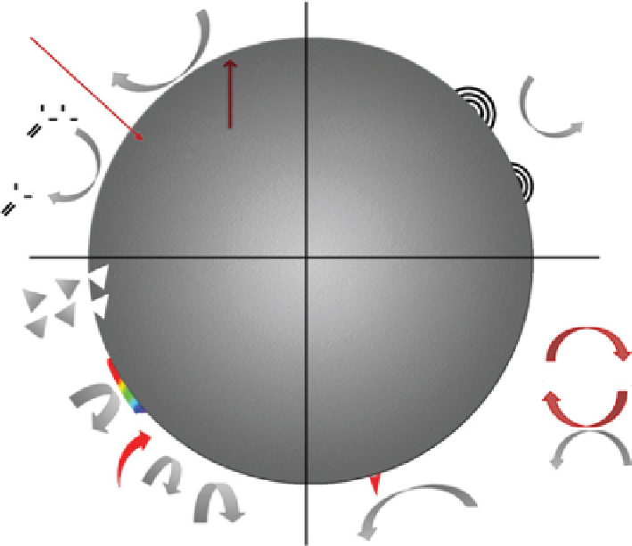
Although apparently we may have a good knowledge of an NM's chemical and physical characteristics, there is a lack of
understanding of the intracellular activity and impact of engineered NMs on cell function. They may interact with a single cell
in different ways than a tissue or whole organism, determining that simple
in vivo
models may not be suitable for complete
interpretation. They may also coat their surfaces, such as, proteins, antibodies, and small biomolecules, depending on the type
of biological fluid they are in contact with (blood, plasma, interstitial fluid), avoiding the immune system. They may even affect
intracellular responses, inducing damage or beneficial responses. Nanostructured metal oxides, for example, may generate
reactive oxygen species such as singlet oxygen, superoxide, and peroxide, as well as participating in oxidation-reduction
processes on the cell surface, which may degrade cell membranes, proteins, and even dNA. Interaction with biomolecules may
also induce changes in their functional structures or block the active sites of enzymes, which in turn will not always have good
metabolic consequences [3] (Fig. 30.6).
In econanotoxicology, it is important to understand how NMs will interact with a living organism from the moment they are
exposed to it till their degradation or elimination, as well as whether these materials (or their by-products) are bioacummulated
within cells, tissues, and organs, thus inducing intracellular changes, inflammatory responses, or undesirable effects culmi-
nating in metabolic illness. Because nanotoxicology is a new research topic of interest, there are many contributions as attempts
to standardize the evaluation of NM toxicity, considering that the interaction of these materials with dying agents, dNA, and
cellular structures could cause some variability in data interpretation and must be validated carefully [27, 28].
Ecotoxicity tests are tools used within environmental hazard assessment frameworks to answer questions about the intrinsic
dangers of chemical substances that may be released into the environment [29]. These tools can be applied to NMs when they
are evaluated, and the exposure scenarios should be replicated using
in vitro
and
in vivo
toxicity assays to know the potential
O
2
Electron-donor/acceptor
groups
UV
O
-
O
2
e-
HO
OH
CR
O
-
h+
C
Material
composition
e
-
O
UV-activation
HO
R
C
O
Dissolution
Media interactions
Redox cycling and
catalytic chemistry
e
-
Coating may protect
the surface, change
cellular uptake or lead to
release of toxic chemicals
Q
-
Q
Passivation
O
-
O
2
H
2
O
Fe
++
H
2
O
H
2
O
2
Q=quinone
Q
-
= semiquinone
Hydrophobicity: interactions with
cell membranes, determines uptake.
Hydrophilicity: water suspendability.
H
2
O
OH
figurE 30.6
Possible mechanisms by which nanoparticles interact with biological tissue. Adapted from Ref. [3].