Environmental Engineering Reference
In-Depth Information
(a)
(b)
(c)
1 μm
1 μm
1 μm
20
16
12
8
4
0
24
20
16
12
8
4
0
0.25
0.50.751.001.251.50
Position [μm]
1.75
2.00
2.25
2.50
2.75 3.00
0.25
0.5 .751.001.251.50
Position [μm]
1.75
2.00
2.25
2.50
2.75
3.00
0.4
0.8
1.20
1.60
2.00
Position [μm]
1.0
13.5 nm
13.5nm
11.0nm
1.8
0.8
1.6
1.4
1.2
1.0
0.8
0.6
0.4
0.2
3.0 nm
0.6
11.0 nm
0.4
0.2
-5
0
5
10
15
20
25
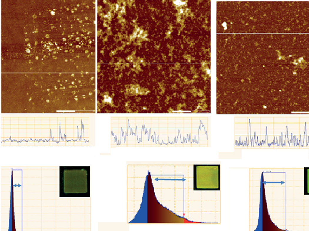
Figure 29.13
Height scanning force microscopy (SFm) images of the glass surfaces modified with the alkoxysilanized humic substances
from different sources. (a) cHP-APTS-100-treated surface with an RmS roughness of about 1.5 nm; (b) PHS-APTS-100-treated surface with
an RmS roughness of 5.9 nm; and (c) AHS-APTS-100-treated surface with an RmS roughness of 3.8 nm. The roughness was determined for
a 3 × 3 µm
2
surface area. For claritym we also include the fluorescence image of the same sample. The graphs below each image represent a
cross section profile along the horizontal line as indicated, and the corresponding height distributions. Reproduced with permission from
Ref. [47]. © Royal Society of chemistry.
At the micron scale (resolution of clm), results show silanized HS producing somewhat homogeneous coatings on DNA
array glass. At the same time, substantial differences could be seen from the fluorescence of HS coatings depending on the
origin of parent materials (peat, coal, and water) modified with 3-aminopropyltrimethoxysilane (APTS). The highest fluores-
cence intensities were observed for peat HS (PHS-APTS-100) and aquatic HS (AHS-APTS-100), which may vary with the
thickness of the self-assembled layer or the amount of attached humic material. The results indicate that peat humic materials
produced the thickest layer. Of particular note is the lack of fluorescence over surfaces treated with parental humic materials.
Nonmodified humic materials, as expected, did not attach to the glass surface due to a lack of positively charged vacancies at
the water-solid interface, which drives their interaction with alumosilicates in nature. The function of positive vacancies was
successfully replaced in this study by incorporating alkoxysilyl groups as anchoring moieties into the humic structure.
The finer-scale structure of these humic coatings was studied on the nanometer scale using the AFm technique. The results
are given in Figure 29.13. AFm images show the surface topography of all three samples—coal, peat, and aquatic humic mate-
rials, immobilized on a DNA glass slide under aqueous conditions. All images are accompanied by the corresponding height
distributions.