Environmental Engineering Reference
In-Depth Information
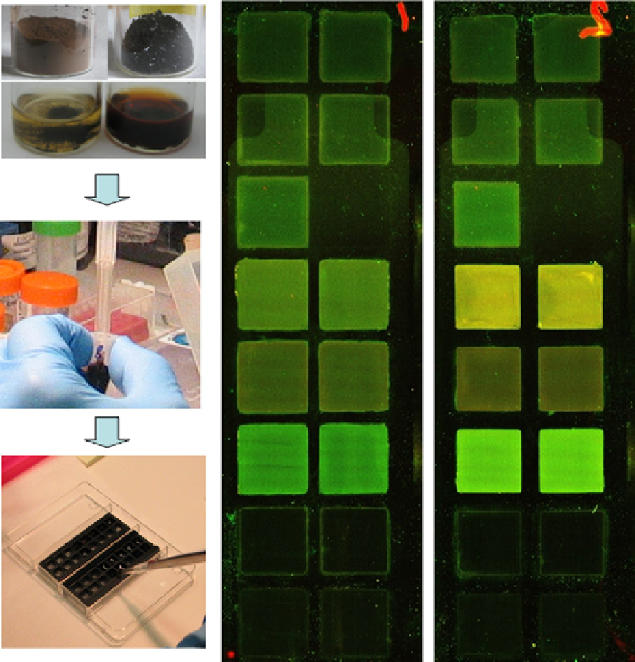
Figure 29.12
General view of the alkoxysilanized humic probes in the solid and dissolved states applied on the surface of DNA array
glass slides that are segregated into multiple patches by the plastic chamber (left part) and the “artificial” color images of the humic adlayers.
The latter are self-assembled in situ at the water/solid interface (right part). The materials in four glass beakers show humic probes made from
coal materials (darkly colored) and peat materials (lighter color). The slides were scanned in a laser microarray scanner at two channels:
ex 532 nm, em 570-590 nm (green); and ex 635 nm, em 680-700 nm (red). The samples are listed from the bottom and are as follows: the
bottom (first) row is a solution of a nonmodified sample of coal humics (cHP); the second row is cHP-GPTS-100; the third row is AHS-
APTS-100; the fourth row is cHP-APTS-100; the fifth row is PHA-APTS-100; the sixth row is a buffer solution (right side, the background
control); and cHP-APTS-20 nonheated (left side); the seventh row is cHP-APTS-20 nonheated; the eighth row is cHP-APTS-20 (heated).
Reproduced with permission from Ref. [47]. © Royal Society of chemistry.
of the humic coatings formed as a result of the self-assembly of the silanized humic derivatives into adlayers at the water-
solid interface were studied using confocal laser microscopy (clm) and atomic force microscopy (AFm) [47]. This was
achieved by preparing humic coatings suitable for AFm studies using DNA array slides as solid supports. The immobili-
zation procedure for the alkoxysilyl humic derivatives and the clm images of the resultant coatings are shown in
Figure 29.12.