Environmental Engineering Reference
In-Depth Information
of the fibrous structure of CNTs [68, 70]. The forces responsible for bacterial adsorption can be van der Waals forces, electrostatic
interaction, and hydrophobic interaction [68, 71-73]. The bacterial cells that have direct contact with CNTs upon adsorption
have a strong tendency to form biofilms or aggregates by connecting individual cells. The adsorption of bacteria onto CNTs
may occur through the following steps: (i) diffusion of bacteria from a solution to the outer surface of CNTs; (ii) diffusion of
bacteria into the macropores of CNTs; (iii) adsorption of bacteria onto the internal surface of CNTs; and (iv) formation of
biofilm or aggregates on the CNT surface [67].
The main reason for using CNTs as adsorbents apart from providing adsorption sites to capture bacteria is the antimicrobial
property of CNTs, which is a unique feature. The antimicrobial property enables CNTs to inhibit bacterial cell activity, causing
cell inactivation. The antimicrobial property of CNTs can be attributed to the impairment of cell function through the destruc-
tion of major constituents (for instance, the cell wall), interference with cell metabolic processes, and inhibition of cell growth
by blocking the key cellular constituent (for instance, DNA, coenzymes, and cell wall proteins) [74]. Bacterial cells, especially
those in direct contact with CNTs, may immediately lose their cellular integrity after exposure to CNTs. The antimicrobial
activity of CNTs is significantly influenced by time. A longer exposure time is known to increase CNT antibacterial activity and
increase cell inactivation. The increased activity is due to the increased probability of contact between CNTs and bacterial cells,
which diminishes the resistance of bacteria after a long exposure time [75, 76]. Two types of bacteria exist based on their cell
wall structure: Gram-positive and Gram-negative. The cell wall of Gram-positive bacteria is usually composed of a thicker
peptidoglycan layer that is susceptible to attack by CNTs. The cell wall of Gram-negative bacteria comprises a thin peptido-
glycan layer and an additional outer layer composed of phospholipids and lipopolysaccharides. CNTs reportedly possess
stronger antimicrobial activity in Gram-positive bacteria than in Gram-negative bacteria. Therefore, the difference in cell wall
structure can affect the antimicrobial activity of CNTs [77].
The nanoparticle size is also another factor that affects the antimicrobial activity of CNTs. Kang et al. [78] compared the
antimicrobial activities of SWCNTs and mWCNTs toward
E. coli
and found that bacterial cell damage was much more pro-
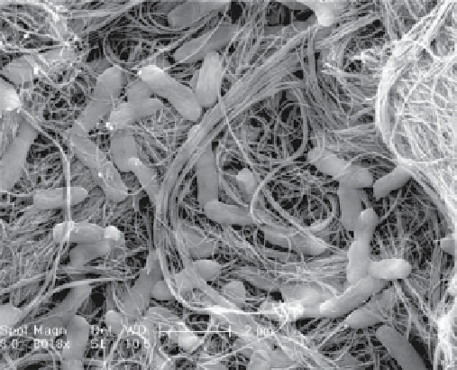
nounced in SWCNTs than in mWCNTs. Scanning electron microscopy (SEm) images of
E. coli
after exposure to SWCNTs
and mWCNTs are shown in Figure 8.2. The result implies that the smaller diameter and shorter length of SWCNTs benefit
antimicrobial activity by providing a larger surface area to increase the likelihood of CNTs making contact with bacterial cells.
The smaller diameter of SWCNTs also facilitates the partitioning and partial penetration of CNTs into the cell wall. Arias et al.
[77] reported stronger interactions in SWCNTs and observed that SWCNTs have tight contact with bacterial cells. These
researchers also believed that the needlelike appearances of SWCNTs surrounding cell aggregates are more likely to inflict
severe cell damage. Bacterial cells make only loose contact with mWCNTs by settling on the mWCNT surface, which may not
effectively damage cell walls, unlike SWCNTs.
SWCNTs demonstrate higher affinity for interacting with bacterial cells than mWCNTs, but SWCNTs involve high synthesis
costs. Therefore, mWCNTs are modified to enhance their performance. Yuan et al. [79] grafted mWCNTs with polyamidoamine
(PAmAm) and deposited silver nanoparticles. The presence of PAmAm debundles mWCNTs, enhancing dispersion. Silver
nanoparticles significantly improve the antimicrobial efficiency of mWCNTs against
S. aureus
. Zardini et al. [80] functionalized
(a)
(b)
fiGure 8.2
Scanning electron microscopy (SEm) images of
E. coli
after exposure to (a) mWCNTs and (b) SWCNTs [78].