Biology Reference
In-Depth Information
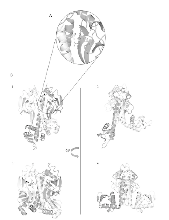
Figure 1.3 Comparison between homo-dimers of unliganded RrCooA (PDB: 1FT9) and
imidazole bound ChCooA (PDB: 2FMY) crystal structures. (A) The CooA haem group dis-
plays a unique iron axial coordination. The haem proximal ligand is His77, where the
distal ligand is Pro2 of the other subunit of the dimer. (B) The frontal and side views
of the unliganded RrCooA (1 and 2, respectively) are compared with the frontal and side
views of the imidazole bound ChCooA (3 and 4, respectively). When no ligand is coor-
dinated to the haem-iron atom, the homo-dimer displays an asymmetric conformation
(2). On the other hand, upon ligand binding, structural rearrangements cause move-
ments of the DNA-binding domain, and the homo-dimer acquires a more symmetric
structure (4).