Image Processing Reference
In-Depth Information
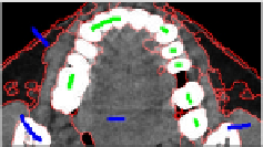
a
b
Fig. 9.12
User interaction and merged blocks. (a) Initial classification and user markers in
2D MPR image (Green and blue markers indicate the object and background respectively).
(b)2DviewofmergedblocksbyBCA.
lines indicate edges between the homogeneous blocks pre-classified by 3D mean
shift. BCA merging method can efficiently separate dental object.
It is worth noting that the initial homogeneous blocks merging is crucial for sub-
sequent learning processing, which can efficiently reduce the dimensions of com-
puting matrix. The proposed segmentation method can be described as a general
cluster-then-label algorithm of semi-supervised classification.
Algorithm:
Cluster-then-Label.
Input:
original voxels data
V
i
∈
Ω
, an unsupervised clustering algorithm
A
, labeled
blocks
(
b
1
,
y
1
)
,···,
(
b
l
,
y
l
)
, unlabeled data
b
l
+
1
,···,
b
l
+
u
, and a semi-supervised
learning algorithm
SSL
.
Output:
labels on unlabeled blocks
y
l
+
1
,···,
y
l
+
u
.
b
l
+
u
using
A
.
2.
For each resulting clustered block, let
S
be the labeled instances. Learn a semi-
supervised predictor from
S
:
f
s
=
1.
Clustered blocks
b
1
,···,
. Apply
f
s
to all unlabeled instances.
In step 1, the clustering algorithm
A
is unsupervised. In step 2, one predictor
is we learned using the labeled instances to apply for all the unlabeled instances.
This cluster-then-label algorithm does not necessarily involve a probabilistic mix-
ture model.
In validation, we first verify the segmentation algorithm based on blocks maxi-
mum similarity (BMSRM) derived from traditional 2D MSRM, and then implement
the BCA strategy on the same volume data as a comparison. The semi-supervised
learning is used to adjust classification errors, and finally provide the accurate object
contour. The program is implemented on a PC (Intel Xeon E5620 2.4 GHz CPU,
24GB RAM, VC++ 9.0 programming environment). The proposed BCA scheme
is relying only on simple neighborhood nonlinearities, no optimization or post-
processing is required. Figure 9.13 demonstrates a CT sequence (512
SSL
(
S
)
100)
with resolution of [0.94, 0.94, 2.0] mm. The first row lists the interaction in MPR
planes after initial classification; the second row lists the corresponding 2D view of
segmentation results of BCA. The green marker represents the dental object while
the blue markers represent the background in the selected axial, sagittal and coronal
image planes. The marker blocks cover only part but representative features of de-
sired dental objects. The total computing time of the proposed BCA method takes
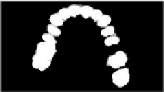
about 2.63 seconds on the whole CT data. The extracted teeth models are listed in
Figure 9.14. Another important issue is that the local resulting errors revision needs
×
512
×

Search WWH ::

Custom Search