Geology Reference
In-Depth Information
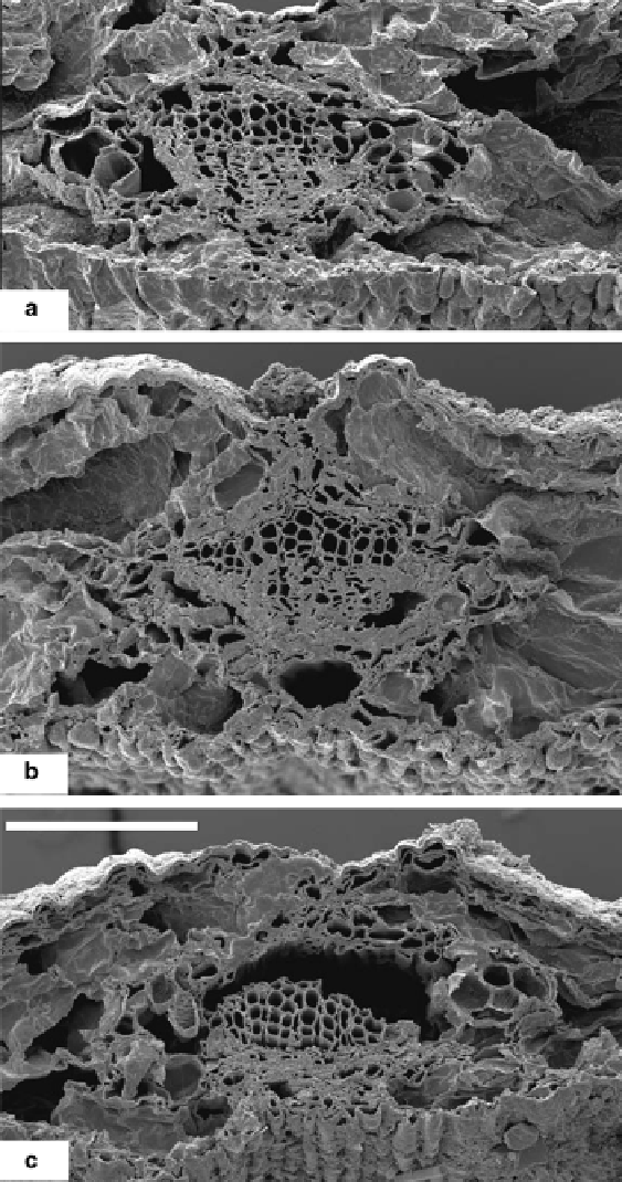
Fig. 1.4
SEM photographs of transverse section of leaves of decay series samples of
Metasequoia
glyptostroboides
, showing only the midvein portion, with adaxial surface upwards. (
a)
Sample 1.
(
b)
Sample 5. (
c
) Sample 6. Compared with Sample 1 (
a
), cells of Sample 5 (
b
) have not changed
much but those of Sample 6 (
c
) largely collapsed. Scale bar 100
μ
m (for all three fi gures.)
Search WWH ::

Custom Search