Geology Reference
In-Depth Information
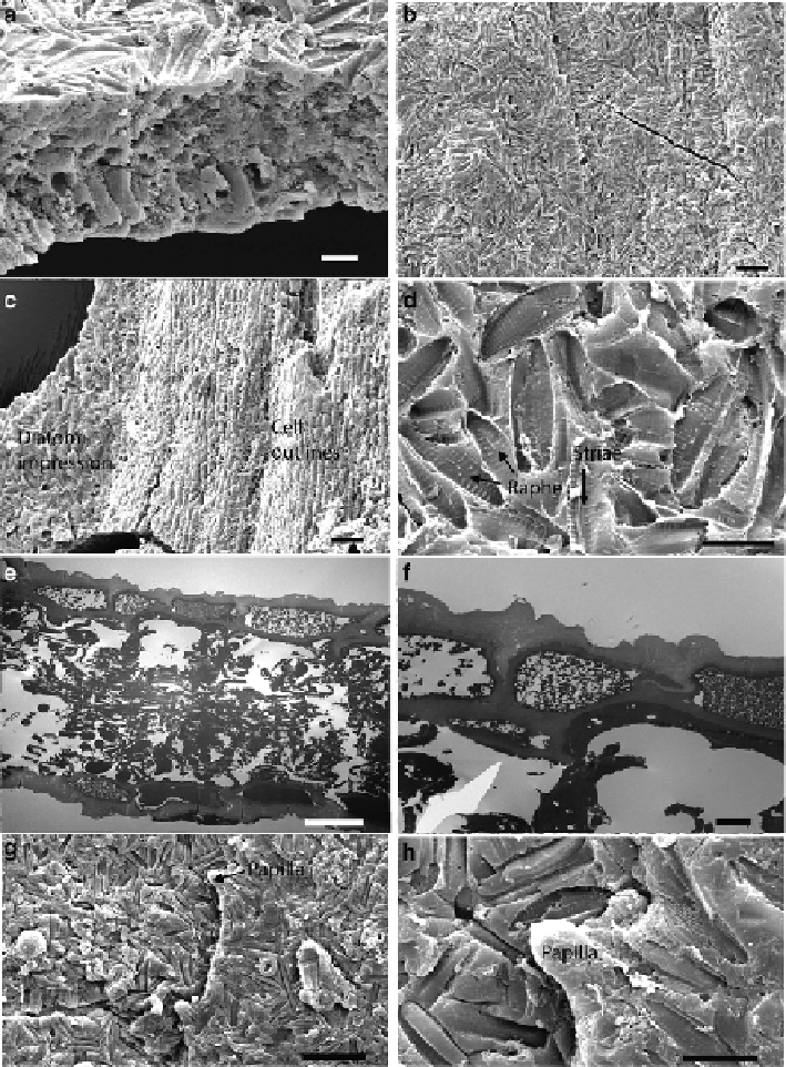
Fig. 4.2
Scanning and transmission electron microscopy of fossil plants from Enspel. (
a
-
d
)
Dicotyledon leaf, (
e
-
i
) conifer leaf. (
a
) Fractured edge showing thickness of organic matter and
possible internal cellular organisation. (
b
) Surface covered in diatom impressions. (
c
) Diatom
impressions and probable epidermal cell outlines where diatoms are absent. (
d
) Details of diatom
impressions showing their complex, sometimes overlapping arrangement. (
e
) Entire cross-section
of fossil conifer showing epidermis and epidermal cells on both surfaces and remnant internal
organisation. (
f
,
g
) Details of the epidermis with cuticle. The cuticle is the thick, outer, more elec-
tron lucent (i.e., paler) layer showing details of microlaminated cuticle ultrastructure in (
g
). (
h
,
i
)
Leaf margin papillae and conifer surface obscured by diatoms. Scale bars: 10 lm in (
a
), (
d
), (
e
), (
i
);
Search WWH ::

Custom Search