Environmental Engineering Reference
In-Depth Information
Dex
(a)
Dex
HSP
90
HSP
90
GR
HSP
90
HSP
70
HSP
70
GR
HSP
70
HSP
70
HSP
90
GR
Dex
Cytoplasm
Dex
Dex
GR GR
Nucleus
Dex Dex
Basal
complex
GR GR
(b)
Hormone
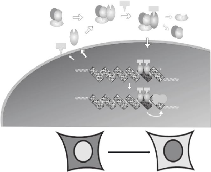
FIGURE 35.2
(See color insert.)
Glucocorticoid receptor (GR) biology. (a) At resting state, GR resides in the cytoplasm as a
part of a multiprotein complex with heat-shock proteins and immunophilins. (From Pratt, W.B. and D.O. Toft,
Endocr. Rev.
, 18, 306, 1997.) Upon hormone stimulation, GR dissociates from this complex and migrates to the
nucleus (cytoplasmic-to-nuclear translocation). In the nucleus, the receptor interacts with GR regulatory ele-
ments (GREs) throughout the genome, and regulates GR-mediated transcriptional responses. (b) Schematic
representation of the translocation of the green luorescent protein (GFP)-tagged GR from the cytoplasm to the
nucleus in the presence of hormone or EDCs with glucocorticoid activity. This translocation is the basis of the
biological screening for detection of water contaminants interacting with a speciic nuclear receptor(s).
(Figure 35.2a), where it interacts with GR genomic regulatory elements (GREs) to elicit
hormone-speciic transcription regulation (John et al., 2008).
Similarly to the GR, AR is largely cytoplasmic in the absence of its ligand, and rapidly
translocates to the nucleus in response to testosterone (Klokk et al., 2007). We used cell
lines engineered to express GFP-tagged GR and AR constructs under a tetracycline-
repressible promoter (Walker et al., 1999; Klokk et al., 2007). In these cells, tetracycline
suppresses the expression of the receptors, and removal of the drug results in protein
expression. When cells are plated on coverslips overnight in a medium supplemented with
hormone-free (charcoal-stripped) serum without tetracycline, cytoplasmic expression of
GFP-tagged receptors can be readily observed, and translocation to the nucleus occurs
in 30 min (Figure 35.2b). Upon exposure to corticosteroids (such as dexamethasone) or a
vehicle control. Cells are ixed, counterstained with 4′,6-diamidino-2-phenylindole (DAPI)
for visualization of the nuclei, and translocation is visualized on a luorescent microscope.
Images are acquired in the green (GFP-GR and GFP-AR) and UV (DAPI) channel, and
receptor translocation is scored as “negative,” “partial,” or “complete” (representative neg-
ative and complete translocation of the GFP-GR are shown in Figure 35.3a).
We further developed an automated, quantitative high throughput read-out of the
translocation assay using a Perkin Elmer OPERA imaging system (Figure 35.3b). For these

